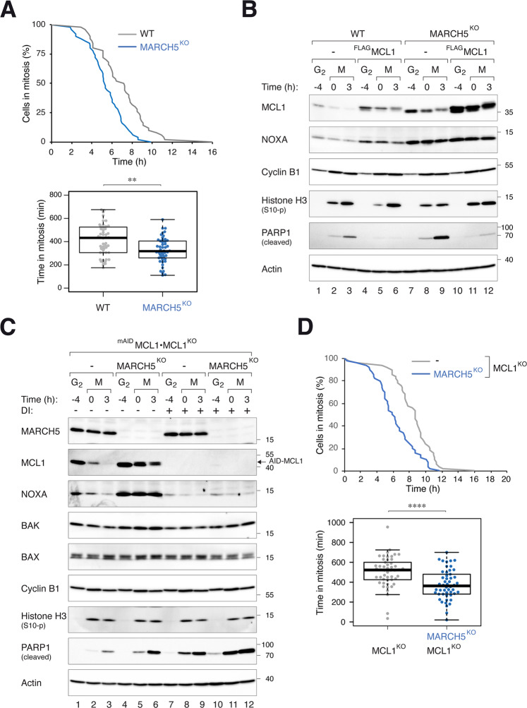
Fig. 3. MARCH5 inhibits mitotic apoptosis through MCL1-dependent and -independent mechanisms.
A Depletion of MARCH5 accelerates mitotic apoptosis. WT and MARCH5KO cells were transfected with histone H2B-GFP before synchronized and arrested in mitosis as before. Individual cells were tracked using live-cell imaging for 24 h (starting at 6 h after second thymidine release) (n = 50). The duration of mitotic arrest is plotted using Kaplan-Meier estimator. Box-and-whisker plots show the elapsed time between mitotic entry and mitotic apoptosis/slippage. **p < 0.01. B Ectopic expression of MCL1 can abolish MARCH5KO-mediated mitotic apoptosis. Cells expressing FLAGMCL1 were generated in HeLa or MARCH5KO backgrounds. The cells were synchronized and arrested in mitosis as before. Protein expression was analyzed with immunoblotting. C Disruption of MCL1 promotes more apoptosis in MARCH5KO cells. MARCH5 was inactivated with CRISPR-Cas9 in MCL1KO expressing mAIDMCL1. The cells were synchronized and arrested in mitosis as before. mAIDMCL1 was turned off with DI. Protein expression was analyzed with immunoblotting. D Additive acceleration of mitotic apoptosis in the absence of MCL1 and MARCH5. MCL1KO cells expressing mAIDMCL1 in either WT or MARCH5KO background were transiently transfected with histone H2B-GFP before synchronized and arrested in mitosis as before. The cells were incubated with DI (to turn off MCL1) before individual cells were tracked using live-cell imaging for 24 h. The duration of mitotic arrest is plotted using Kaplan–Meier estimator. Box-and-whisker plots show the elapsed time between mitotic entry and mitotic apoptosis/slippage. ****p < 0.0001.

