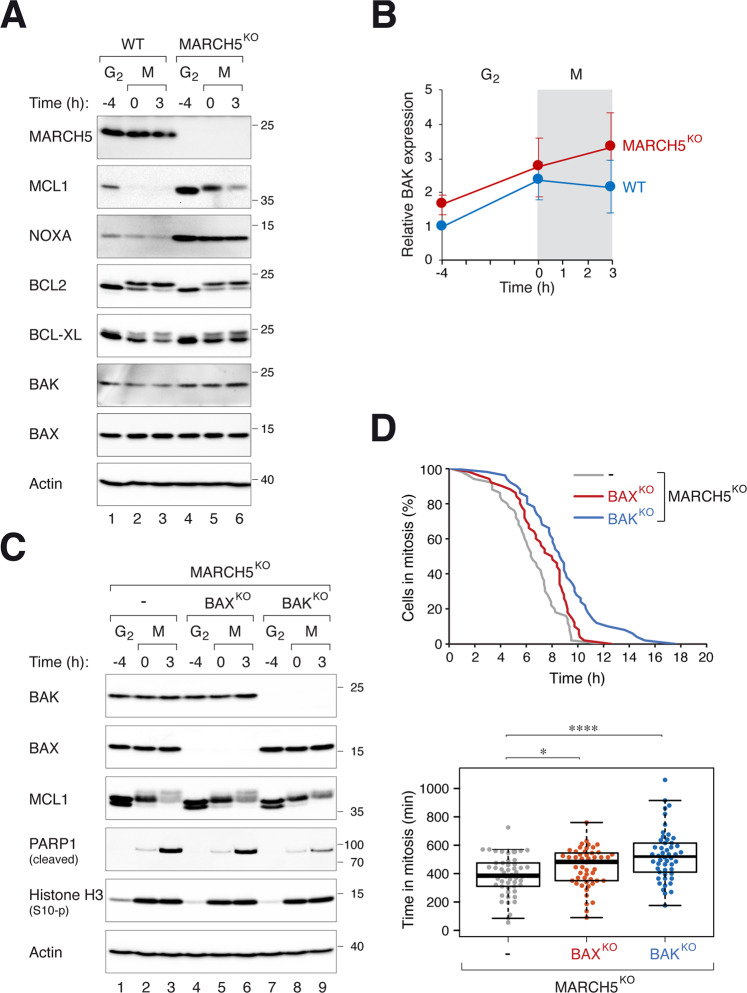
Fig. 5. MARCH5 regulates mitotic apoptosis through BAK.
A Disruption of MARCH5 elevates BAK expression. WT and MARCH5KO cells were synchronized and arrested in mitosis as before. The expression of the indicated proteins was analyzed with immunoblotting. Note that several proteins (MCL1, BCL2, and BCL-XL) displayed mitotic gel mobility shifts. B Increased expression of BAK during mitotic arrest in MARCH5KO. WT or MARCHKO cells were synchronized and arrested in mitosis as before. The expression of BAK during G2 and mitosis was quantified from immunoblots of three independent experiments (mean ± SEM). C PARP1 cleavage in MARCHKO-mediated mitotic apoptosis is reduced in the absence of BAK. BAX or BAK was ablated with CRISPR-Cas9 in MARCH5KO cells. The cells were synchronized and arrested in mitosis as before. Protein expression was analyzed with immunoblotting. D MARCHKO-mediated mitotic apoptosis is delayed in the absence of BAK. MARCH5KO, MARCH5KOBAXKO, and MARCH5KOBAKKO (all were MCL1KO cells expressing mAIDMCL1) were transiently transfected with histone H2B-GFP before synchronized and arrested in mitosis as before. Individual cells were then tracked using live-cell imaging. The duration of mitotic arrest is plotted using Kaplan–Meier estimator. Box-and-whisker plots show the elapsed time between mitotic entry and mitotic apoptosis/slippage. *p < 0.05; ****p < 0.0001.

