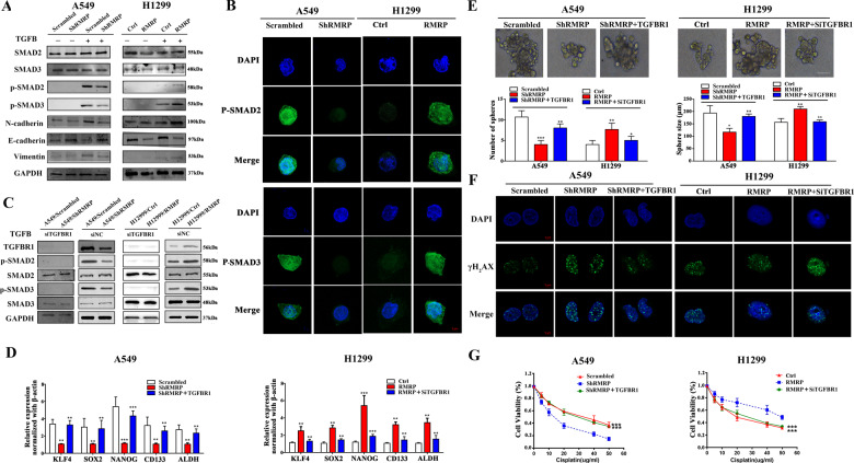
Fig. 7. RMRP regulated the TGFBR1/SMAD2/ SMAD3 pathway in NSCLC.
A A549/Scrambled, A549/shRMRP, H1299/Ctrl, and H1299/RMRP cells under the TGFB treatment were collected and subjected to Western blot analysis using the indicated antibodies. The TGFB treatment was stimulated with 5 ng/mL TGFB for 1 h (n = 3). B Representative immunofluorescence images of anti-p-SMAD2/3 (green) and DAPI (blue) staining of the treated NSCLC cells. The cells were also cultured under the TGFB treatment and were collected for immunofluorescence analysis (n = 3) (Scale bar, 5 μm). C A549/Scrambled, A549/shRMRP, H1299/Ctrl, and H1299/RMRP cells were treated with TGFBR1 knockdown (siTGFBR1) or siRNA negative controls (siNC). The samples under the TGFB treatment were collected and subjected to Western blot analysis using the indicated antibodies (n = 3). D Related cells were collected and analyzed by qRT-PCR (n = 3). β-actin was used as the reference for normalization. E The number of tumorspheres was counted, and the morphology was observed under a light microscope. Upper panel: Representative pictures of tumorspheres. Lower panel: Tumorspheres quantification results including the number and size of spheres. (Scale bar, 100 μm). F The treated A549 and H1299 cells were subjected to irradiation (6 Gy). Subsequently, immunofluorescence analysis was performed after 1 h (n = 3, Scale bar, 5 μm). G Results of CCK-8 assays showing cell viability after incubation with different concentrations of cisplatin for 24 h. *P < 0.05; **P < 0.01; ***P < 0.001. Data represent three independent experiments.

