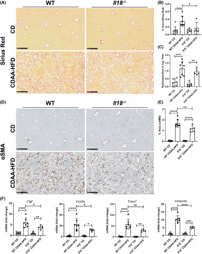
FIGURE 5.

IL‐18 deficiency reduced CDAA‐HFD–induced fibrosis. (A, B) Collagen deposition assessed by Sirius Red staining and (C) hydroxyproline, (D, E) αSMA‐positive cells, and (F) mRNA expression levels of the profibrotic genes Ctgf, Col3a, Timp1, and Vimentin in WT and Il18 −/− mice fed with CDAA‐HFD compared with mice on CD. αSMA, alpha smooth muscle actin; CD, control diet; CDAA‐HFD, choline‐deficient, L‐amino acid‐defined high fat diet; Ctgf, connective tissue growth factor; Col3a, collagen type III alpha 1 chain; Timp1, tissue inhibitor of matrix metalloproteinase 1; WT, wild type. n = 5–8 mice per group for all measured values; scale bars represent 250 μm. *p < 0.05; **p < 0.01; ***p < 0.001; ****p < 0.0001.
