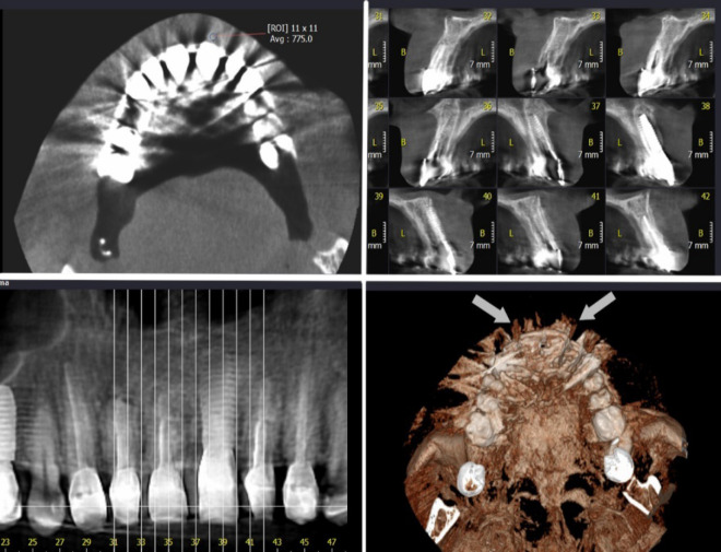
Figure 1.
Axial, cross sectional, panoramic and reconstructed three-dimensional image of maxilla with metallic crowns on the anterior teeth, taken with conventional technique for evaluation after implantation of tooth 9. The metal artifacts can be seen as white opaque lines lining towards buccal and palatal (white arrow in reconstructed image); Note the gray value of the buccal soft tissue

