
Fig. 1.
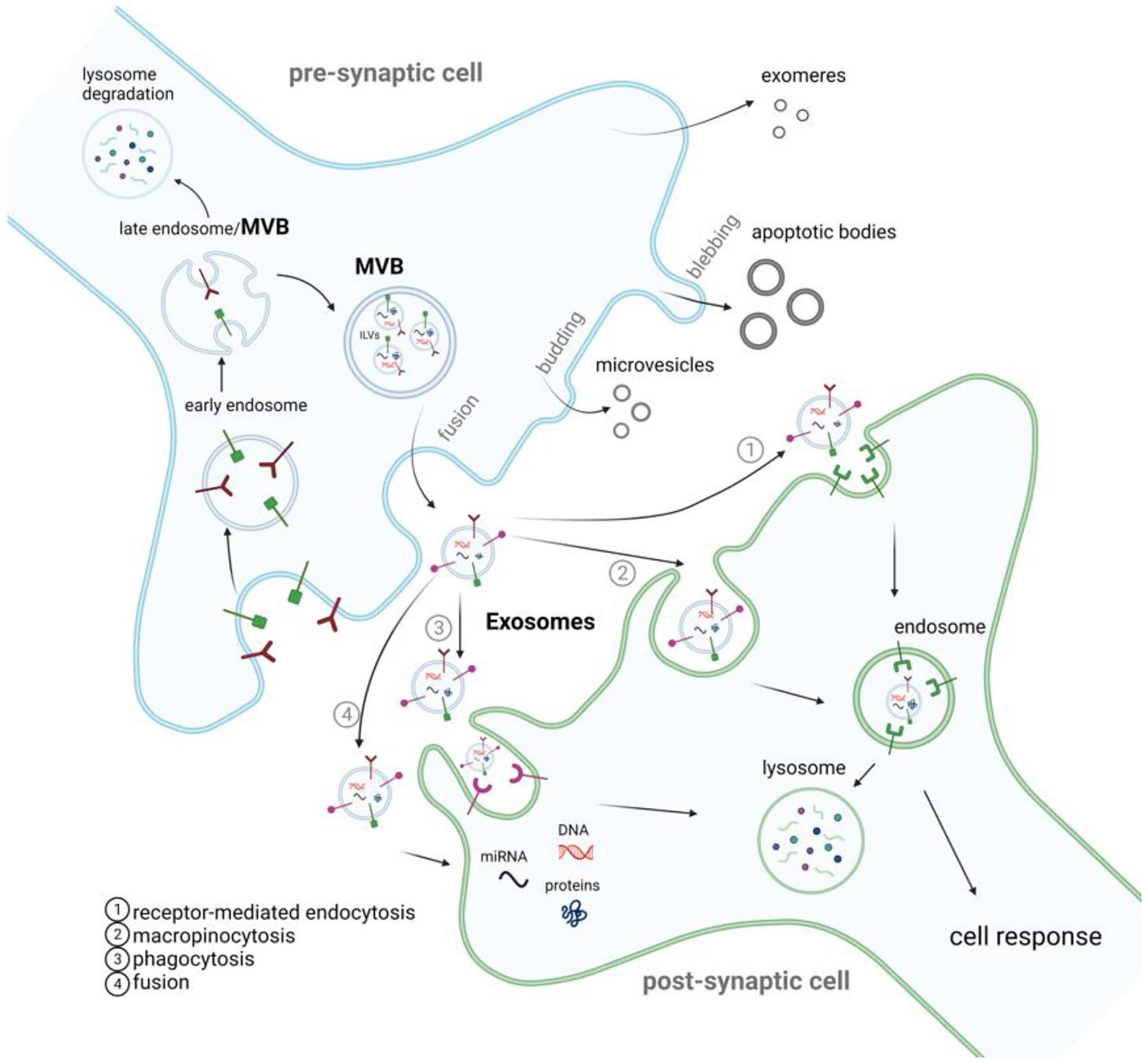
Extracellular vesicle life cycle. Extracellular vesicles (EVs) are membranous nano-sized particles released from the cell and are classically classified into exosomes, microvesicles, and apoptotic bodies. On the donor cell, exosomes derive from the endolysosomal pathway. Following endocytosis, early endosomes are generated and mature into late endosomes that will suffer further internal invagination to form intraluminal vesicles (ILVs). The MVBs fuse with the plasma membrane to release ILVs into the extracellular space as exosomes. Microvesicles are released through membrane budding, while apoptotic bodies are released by dying cells through the blebbing of the plasma membrane. Exomeres are a novel entity whose biogenesis is still being uncovered. Upon EVs release (the majority of studies have focused on small EVs – a mixture of exosomes and microvesicles), they are internalized by the recipient cell through several pathways such as receptor-mediated endocytosis (Dubois et al., 2016), macropinocytosis (Golde, 2022), phagocytosis (Berkowitz et al., 2018) and membrane fusion (Chen et al., 2016). Following their uptake by the recipient cell, EV cargo may undergo clearance or induce a cellular response.