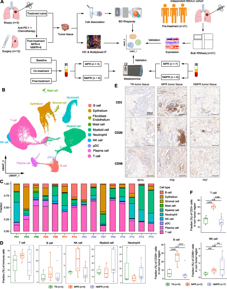
Fig. 1.
scRNA-seq analysis of NSCLC during therapy. A Scheme of the overall study design. B Uniform manifold approximation and projection (UMAP) plot of all cells colored by major cell types according to canonical markers. C Bar plots indicating the proportion of major cell lineages in each patient. D Boxplot showing cellular fractions of T, natural killer (NK), B, myeloid cells and neutrophils in TN (n = 3), MPR (n = 4), and NMPR (n = 8) patients. Center line indicates the median value, lower and upper hinges represent the 25th and 75th percentiles, respectively, and whiskers denote 1.5× interquartile range. Each dot corresponds to one sample. All adjusted P values were larger than 0.05. One-sided unpaired Wilcoxon test was used, and the P values were adjusted by the FDR method. E Representative images of immunohistochemistry (IHC) staining of canonical surface markers for T (CD3), NK (CD56), and B (CD20) cells in a TN (S01b), MPR (P06), and NMPR (P07) patient, respectively. F Quantification of fractions of T, NK, and B cells from the IHC images. One-sided unpaired Wilcoxon test was used

