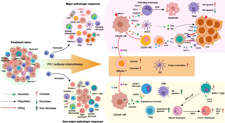
Fig. 8.
Summary of TME dynamics in NSCLC during ICB plus chemotherapy. After ICB plus chemotherapy, the phenotype of immune cells was remodeled, and normal epithelial cells expanded in the TME. The cytotoxic ability of effector T cells was significantly elevated; however, the exhausted markers were also increased. The memory CD8+ T cells were activated into an effector phenotype. Therapy enhanced the antigen-presenting function of DCs. Except these common features, major pathologic responders (MPRs), and non-MPRs had distinct characteristics of TME. The residual cancer cells in MPRs expressed MHC-II molecules to present tumor antigens themselves, and secreted CX3CL1 to recruit PMos, cDC2s, and CD16+ NK cells. PMos secreted CFP to promote apoptosis of cancer cells. Tfhs released CXCL13 to recruit CD20+ B cells and these B cells aggregated in the TME. IFNα and TNF from cDC2s drove the production of FCRL4+FCRL5+ memory B cells. The FCRL4+FCRL5+ memory B cells in turn activated Tfhs by CD86-CD28 and CD40-CD40LG interaction. Then, the activated Tfhs secreted IL21 to enhance release GrB from effector T cells though binding to IL21R. These interactions positively boosted the anti-tumor response. Meanwhile, suppressive Tregs and M2 signature of TAMs were decreased in MPRs. In non-MPRs, aberrant estrogen metabolism caused elevated estradiol in the TME. The TME in non-MPRs was still suppressive, with no decrease of M2 signature of TAMs and increase of VEGFA+ monocytes and suppressive signature of Tregs. In addition, the SPP1+ TAMs and CCL3+ neutrophils interacted with each other to promote expansion of themselves: SPP1+ TAMs secreted SPP1 and IL1B to induce the production of CCL3+ neutrophils, and CCL3+ neutrophils in turn to attract SPP1+ TAMs by CCL3 and CCL4

