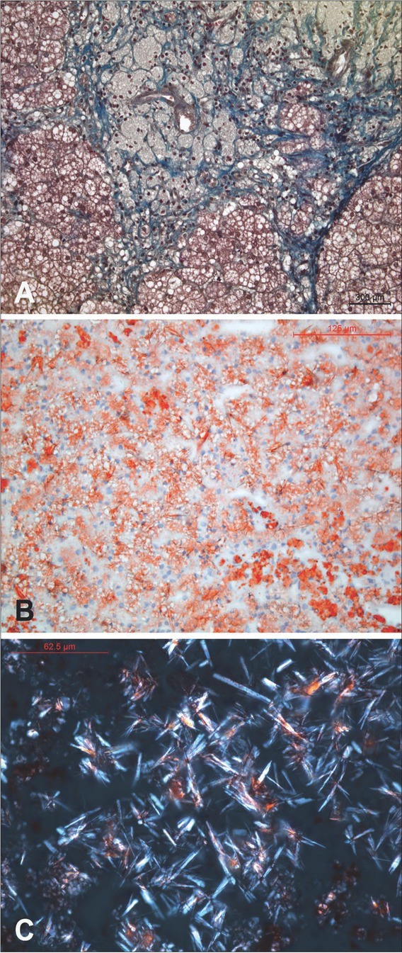
Figure 1.

Liver histology of patient 3. 1A. Foamy macrophages with pyknotic nuclei loaded in the portal space and uniform microvesicular steatosis with accompanying marked fibrosis. (Paraffin section stained with Masson’s trichrome, x200). 1B. Histological picture of liver steatosis with evenly distributed, Sudan IV positive microvesicles in hepatocytes and crystalloid clefts of cholesteryl ester that are only hinted at due to light birefringence. (Frozen section stained with Sudan IV, x25). 1C. Birefringent, anisotropic crystals are seen under polarized light on frozen sections stained by the Sudan IV method. Maltese cross-type birefringence is visible here and there. (Frozen section stained with Sudan IV, polarized light, x40).
