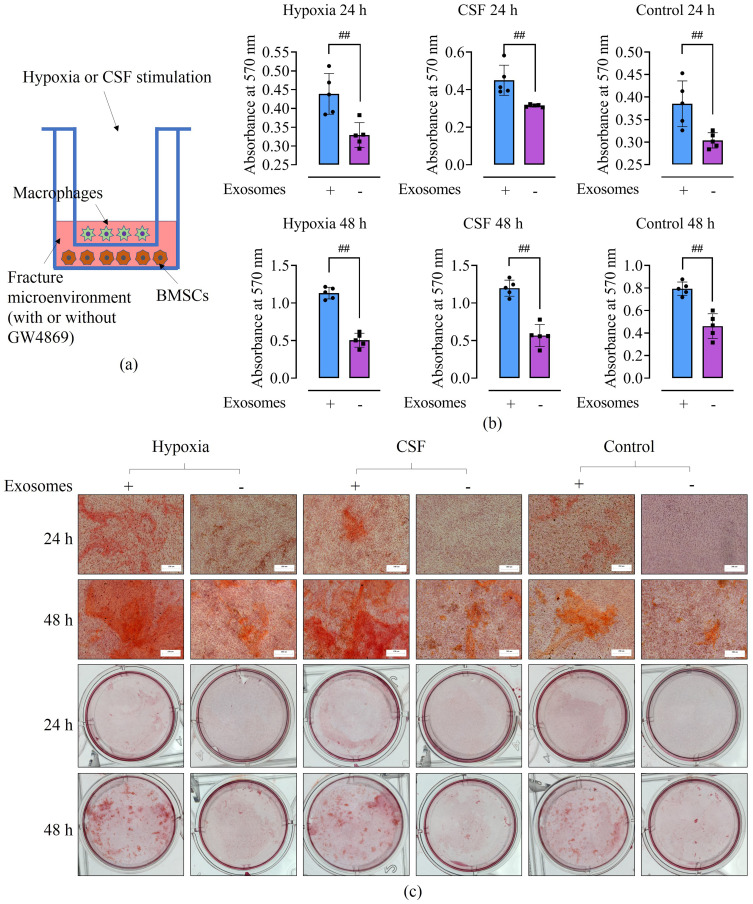
Figure 5.
The effect of exosomes on BMSC osteogenesis in the bone fracture microenvironment. (a) The experimental design; this figure was created using BioRender. (b) The absorbance value of BMSCs at 570 nm after Alizarin red staining. The bar graphs show that in the cell co-culture model, inhibiting the secretion of exosomes by macrophages reduced the OD value of BMSCs after Alizarin red staining. ##p < 0.01. (c) Alizarin red staining of BMSCs, viewed under the microscope and with the naked eye. In the cell co-culture model, inhibition of exosome secretion by macrophages reduced the area of BMSCs stained with Alizarin red, irrespective of whether the macrophages were stimulated using hypoxia, CSF, or BFM alone. Each experiment was repeated five times. Microscopy: 100× magnification and 200 μm scale.

