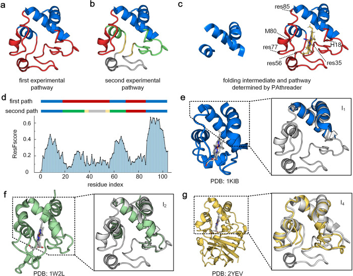
Fig. 5. Folding pathway of horse heart cytochrome c (PDB ID: 1I5T).
a The first experimental pathway. The blue region is first folded and is followed by the red region. b The second experimental pathway. Blue is folded first, followed by green, yellow, red and then grey. It contains 4 intermediates, (blue), (blue + green), (blue + green + yellow) and (blue + green + yellow + red). c Intermediate and folding pathways predicted by PAthreader, the blue region is first folded and is followed by the red region. d Two different experimental paths and the ResFscore distribution of residues identified by PAthreader. e–g Template structures from 1KIB, 1W2L and 2YEV. The solid line box is the partial superposition of templates and the structure of horse heart cytochrome c (grey), which correspond to intermediates of the second experimental pathway.

