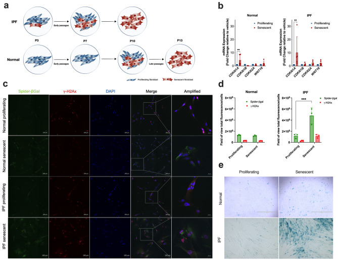
Figure 1.
Lung fibroblasts isolated from normal and IPF patients exhibited a senescent phenotype after underwent through replicative senescence. Schematic presentation of normal and IPF lung fibroblasts transition from early to late passages (a). mRNA expression of senescence associated genes CDKN1A, CDKN1B, CDKN2A, and WNT16 in proliferating and senescent lung fibroblasts from normal or IPF patients (b). Representative images of SA-β-gal and the DNA damage marker γ-H2Ax co-staining is pronounced in senescent lung fibroblasts from normal or IPF patients (c). The scale bars indicate 100 μM (Spider-β-gal, γ-H2Ax, DAPI and merge) and 20 μM for amplified image. Quantification of SA-β-gal and γ-H2Ax total fluorescence/number of cells from proliferating and senescent lung fibroblasts from normal or IPF patients (d). SA-β-galactosidase staining is detected in senescent lung fibroblasts from normal and IPF patients but not in proliferating lung fibroblasts (e). Data are presented as mean ± SD (n = 3 or 5 per group). *p < 0.05, **p < 0.01 and ***p < 0.001 as indicated by the bars.

