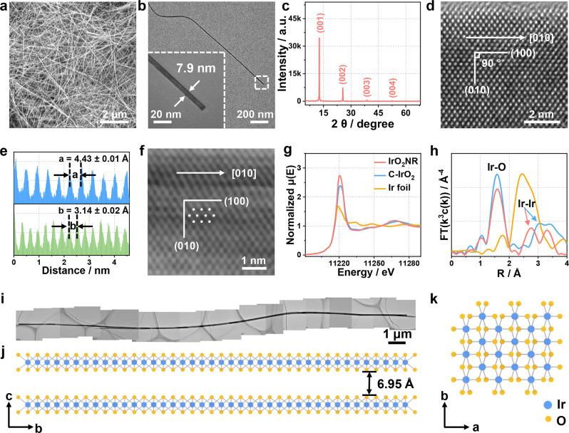
Fig. 1. Morphology and structure of the IrO2NR.
a SEM image showing the uniform distribution of the IrO2NR. b TEM image of a single IrO2NR. The insert is the enlargement of the tail end of the ribbon with a width of 7.9 mm. c XRD pattern. d HRTEM image. e Line scan of the HRTEM image indicated by the (100) and (010) planes. f HAADF–STEM image for the IrO2NR. g XANES spectra and h FT–EXAFS spectra for the IrO2NR, C-IrO2 and Ir foil. i A typical long IrO2NR with a length of 22.53 μm. j, k Pictorial illustration of the crystal structure for the IrO2NR from the (j) a direction and (k) c direction.

