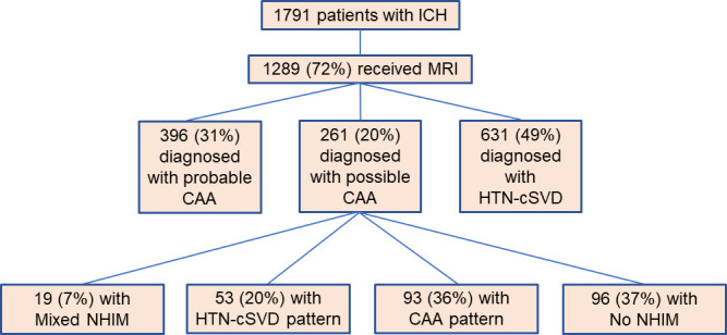
Figure 2.

Participant selection methodology. The above schematic depicts the total number of patients included in the study (n=1791). Of these, 1289 (72%) received an MRI scan of the brain. From this cohort, patients were diagnosed with either probable CAA, possible CAA (isolated lobar ICH) or HTN-cSVD. Lastly, the patients with isolated lobar ICH were further stratified into patients with HTN-cSVD pattern, CAA pattern, mixed NHIM or no NHIM. CAA, cerebral amyloid angiopathy; HTN-cSVD, hyperintensive cerebral amyloid angiopathy; ICH, intracerebral haemorrhage; NHIM, non-haemorrhagic imaging markers.
