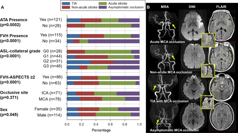
Figure 4.
Symptomatic status comparison between subgroups. (A) Comparison of the proportions of the index events between subgroups of FVH, ATA, ASL-collateral grade, occlusive site and sex. P value indicates results for χ2 test of the proportion of TIA, acute stroke, non-acute stroke and asymptomatic occlusion between groups. (B) Representative cases illustrate the extent of FVH in ipsilateral middle cerebral artery occlusions with acute stroke, non-acute stroke, TIA and asymptomatic status. Arrows indicate FVH. ASL, arterial spin labelling; ASPECTS, Alberta Stroke Program Early Computed Tomography Score; ATA, arterial transit artifact; DWI, diffusion-weighted imaging; FLAIR, fluid-attenuated inversion recovery; FVH, fluid-attenuated inversion recovery vascular hyperintensity; ICA, internal carotid artery; MCA, middle cerebral artery; MRA, MR angiography; TIA, transit ischaemic attack.

