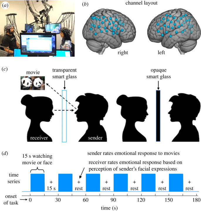
Figure 1.
(a) Set-up for simultaneous neuroimaging of interacting participants separated by a glass panel, i.e. ‘smart glass’ (picture permissions obtained). (b) Channel layout. Right and left hemispheres of a single-rendered brain illustrate median channel locations (blue dots) for 58 channels per participant. Montreal Neurological Institute (MNI) coordinates for each recording channel and corresponding anatomical locations were determined with NIRS-SPM [51]. (c) Paradigm schematic. The ‘smart glass’ divider is transparent during the task (blue bars in (d)) and opaque during rest periods (15 s blank in (d)). (d) Time series for a single run.

