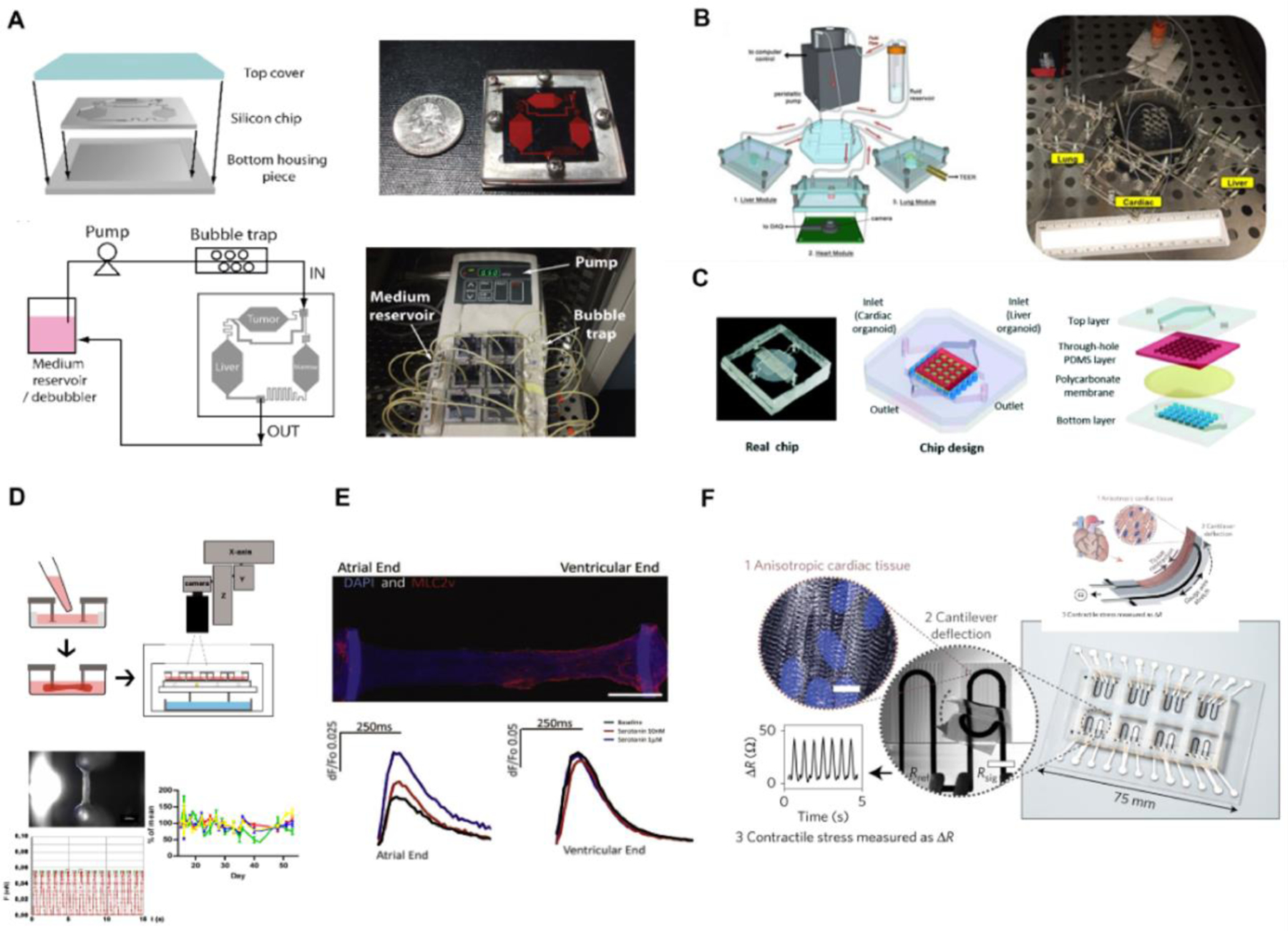
Figure 4: Representative application of organ-on-chip system in drug screening and toxicity testing.

Liver: Panel A-C; Heart: Panel D-F.
(A) Schematics and photographs of a silicon-based perfusable liver-on-a-chip system connected to tumor tissue and bone marrow tissue downstream to study the effect of active metabolite 5-fluorouracil. Reproduced with permission[71]. (B) Schematic and photograph demonstrating a “plug and play” design of liver-on-a-chip hardware system connected to adjacent heart-on-a-chip and lung-on-a-chip systems, demonstrating an altered drug response in heart contraction rate. Reproduced with permission[73]. (C) A design of integrated organ-on-chip system with liver organoid in the top layer and heart tissue organoid at the bottom layer. Metabolism-dependent cardiac toxicity of clomipramine was assessed through a change in heart contraction rate. Reproduced with permission[74]. (D) Schematic overviews of a post-based heart-on-a-chip system for studying heart output through post deflection. Optical video recording of post deflection can generate contraction force, frequency, and relaxation velocity through post geometry and elastic properties. Reproduced with permission[97]. (E) An immunostaining image of heteropolar atrial and ventricular cardiac engineered tissue on Biowire II system. Serotonin only affects the calcium transient to the atrial region of the cardiac construct in a concentration-dependent manner. Reproduced with permission[17]. (F) A schematic and immunostaining image of cardiac muscular thin-film technology with embedded multilayer cantilevers to measure the contraction of an anisotropic cardiac tissue. A soft strain gauge sensor embedded in the PDMS film measures the changes of resistance corresponding to cardiac contractile outputs. Reproduced with permission[7].
