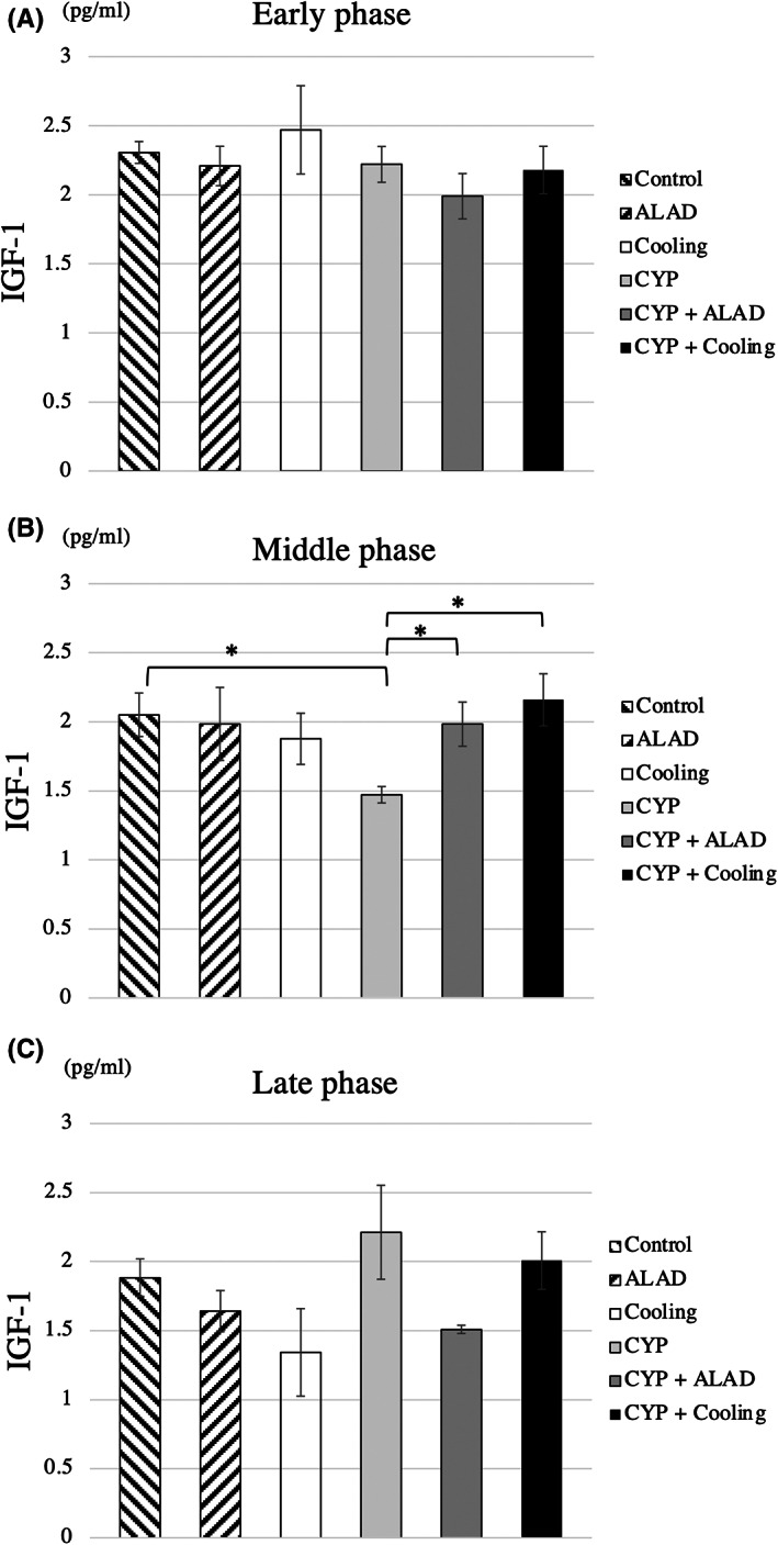
FIGURE 3.

IGF‐1 measurements. Comparison of IGF‐1 in the early (A), middle (B), and late phases (C). Means and standard errors of IGF‐1 are shown as the vertical and the error bars, respectively. Statistical analysis was performed using Student's t‐test (*p < 0.05)
