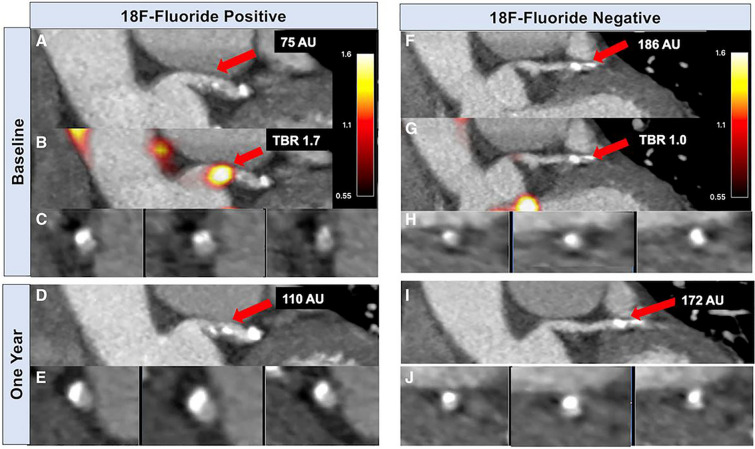
Fig. 3.
Baseline and repeat imaging of two coronary plaques in the same patient. Contrast-enhanced CT coronary angiography (A, C) and fused PET/CT (B) demonstrate NaF activity localized to calcification present in the left main stem. CT coronary angiography 1 year later (D, E) demonstrates increased CT calcification at this site. By contrast, CT coronary angiography (F, H) and PET/CT (G) showed a non-NaF avid calcified plaque in a proximal obtuse marginal branch, which was not found to have increased calcification after 1 year (I, J). Image reprinted without changes from Doris et al. [72] under the Creative Commons Attribution 4.0 International License (CC BY). https://creativecommons.org/licenses/by/4.0/

