FIG. 2.
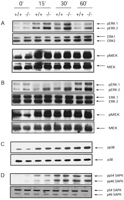
PMA- and TCR-induced ERK activation is increased in spleen cells from HePTP−/− mice. (A) Spleen cells (2 × 10⁷) were unstimulated or stimulated with 10 nM PMA for 15, 30, and 60 min. Activated, phosphorylated ERK, total ERK, activated phosphorylated MEK, and total MEK were detected by immunoblotting. While HePTP+/+ and HePTP−/− splenocytes have similar amounts of total ERK and MEK, HePTP−/− cells demonstrate increased activation of ERK but not MEK after PMA stimulation. (B) TCR stimulation-induced activation of ERK and MEK was evaluated in total splenocytes from wild-type and HePTP deletion mice. Cells were incubated for 0, 15, 30, or 60 min with anti-CD3Ε (10 μg/ml), lysed, and evaluated by immunoblotting for ERK1-2, MEK, phospho-ERK, and phospho-MEK. HePTP−/− lymphocytes demonstrate increased activation of ERK after TCR stimulation compared with that of wild-type controls but no difference in MEK activation. (C and D) p38HOG activation (C) and SAPK activation (D) in HePTP+/+ and HePTP−/− splenocytes. Cells (2 × 10⁷) were treated with sorbitol (400 mM) for the indicated times. Lysates were analyzed by Western blotting for total p38HOG or SAPK and for the phosphorylated activated forms as indicated. Cells from wild-type HePTP and HePTP−/− mice show similar activation profiles for p38HOG and SAPK.

