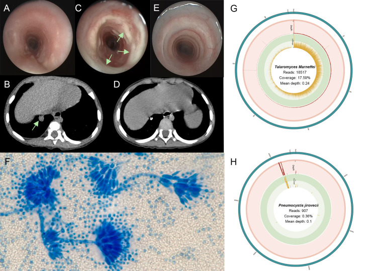
Figure 1.
(A) The first bronchoscope showing a little mucus in the airway. (B) Three-dimensional computed tomography reconstruction of lung window showing a round and high-density shadow in the basal segment (arrows). (C) The bronchoscopic image showing plenty of white secretion in the tracheal inner membrane (arrows). (D) After one year, chest computed tomography showed the nodule shadows was smaller than before, and the calcification was obvious. (E) One year after treatment, tracheoscopy showed no secretion adhesion in the trachea. (F) The lactophenol cotton blue of lavage fluid-stained slide on day 33 showing Talaromyces marneffei with broom-like branches (oil immersion lens, 1000× magnification). (G) T. marneffei coverage and depth in BALF metagenome next-generation sequencing (mNGS). (H) Pneumocystis jirovecii coverage and depth in BALF mNGS.

