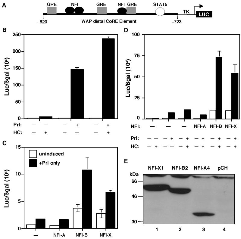
FIG. 3.
Cooperativity of GR and STAT5A on the WAP CoRE. (A) Schematic diagram of the pWAPtk-luciferase construct. (B) GR and STAT5A cooperativity. The pWAPtk-luciferase construct (1 μg) was transiently transfected into JEG-3 cells along with GR (0.2 μg), STAT5A (1 μg), PrlR (0.3 μg), and RSV-β-gal (0.3 μg). Twenty-four hours after induction with hormones (1 μg/ml of both HC and Prl), luciferase expression was measured and transfection efficiencies were normalized to β-galactosidase activity. Cells were induced with HC, Prl, and both HC and Prl, as indicated. (C) Cooperativity of STAT5A and NFI isoforms (0.2 μg, 0.5 μg, and 1 μg of A, B, and X, respectively) for the activation of the WAP CoRE. Three NFI isoforms were transfected into JEG-3 cells along with STAT5A, PrlR, pWAPtk-luciferase, and RSV-β-gal, and the cells were induced with Prl. Cells were transfected with STAT5A and PrlR and induced with Prl and with NFI-A, -B, and -X, respectively, as shown. (D) Cooperativity of GR, STAT5A, and NFI isoforms on the WAP CoRE. Cells were transiently transfected with GR, STAT5A, the PrlR, and pWAPtk-luciferase along with RSV-β-gal. In addition to these constructs, cells were transfected with either NFI-A, -B, or -X. Cells were induced with HC, Prl, or both HC and Prl as indicated. Error bars represent the standard error of three independent determinations. (E) Expression of the three NFI isoforms in JEG-3 cells. Total proteins were isolated from the cells transfected with NFI-X1 (lane 1), NFI-B2 (lane 2), and NFI-A4 (lane 3) and from cells transfected with only the pCH vector (lane 4). These extracts were analyzed on sodium dodecyl sulfate–8% polyacrylamide gel electrophoresis (SDS–8% PAGE), and Western blots were performed using an anti-HA antibody. The molecular sizes of the protein markers are shown.

