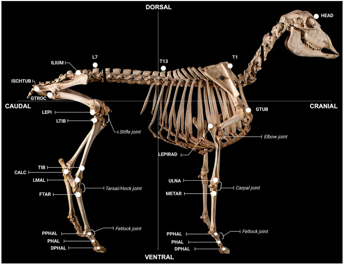
Figure 2.
Anatomical locations for gait assessment. Locations were defined as follows; global: HEAD (on the head between the eyes), T1 (spinous process of T1), T13 (spinous process of T13), L7 (spinous process of L7); forelimb: GTUB, greater tubercle of the humerus; LEPIRAD, lateral epicondyle of the radius; ULNA, distal tubercule of the ulna; METAR, proximal tubercule of the metatarsal; PPHAL, forelimb proximal phalange; PHAL, forelimb phalange; DPHAL, forelimb distal phalange; hindlimb: ILIUM, iliac crest; ISCHTUB, ischial tuberosity; GTROC, greater trochanter of the femur; LEPI, lateral epicondyle of the femur; LTIB, lateral condyle of the tibia; TIB, tibia; CALC, calcaneus; LMAL, lateral malleolus; FTAR, fused tarsal of the metacarpus; PPHAL, hindlimb proximal phalange; PHAL, hindlimb phalange; DPHAL, hindlimb distal phalange. The METAR/FTAR, PPHAL, and PHAL were used to determine the fetlock angle of the fore and hind limbs respectively. The LEPIRAD, ULNA, METAR, and PPHAL and the TIB, LMAL, FTAR and PPHAL, were used to determine the angle of the carpus and tarsus, respectively. The GTUB, LEPIRAD, and ULNA and the GTROC, LEPI, LTIB, and LMAL were used to determine the angle of the elbow and stifle, respectively. Retro-reflective markers attached to each location were 15 mm in diameter, except for markers on the fore- and hind-limb PHAL, PHAL and DPHAL, which were 9 mm. Figure adapted from a sheep skeleton on display at the Museum of Veterinary Anatomy, Faculty of Veterinary Medicine and Animal Science, University of São Paulo, Brazil.

