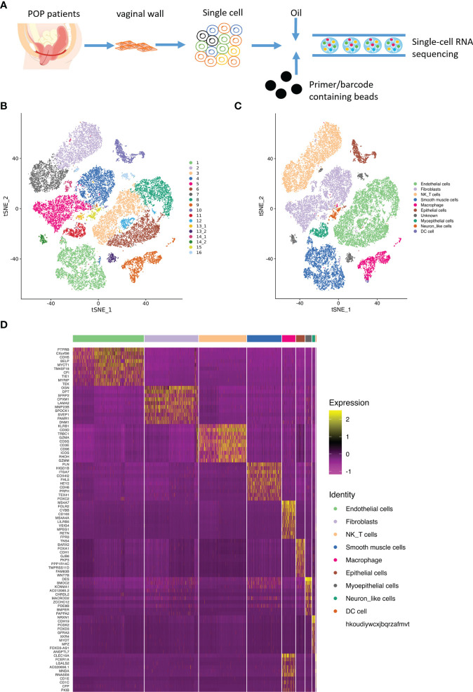
Figure 1.
Main cell lineages in vaginal wall cells. (A). Schematic of tissue dissociation, cell isolation, sequencing, and downstream bioinformatics analysis. (B) tSNE plots of the major vaginal wall cell populations. Each point depicts a single cell, colored according to cell population. (C) Definition atlas of vaginal wall cells (endothelial cells, fibroblasts, NK T cell, smooth muscle cells, macrophages, epithelial cells, myoepithelial cells, Neuron like cell, and DC cell). (D) Heatmap showing the relative expression of marker genes in each cell type.

