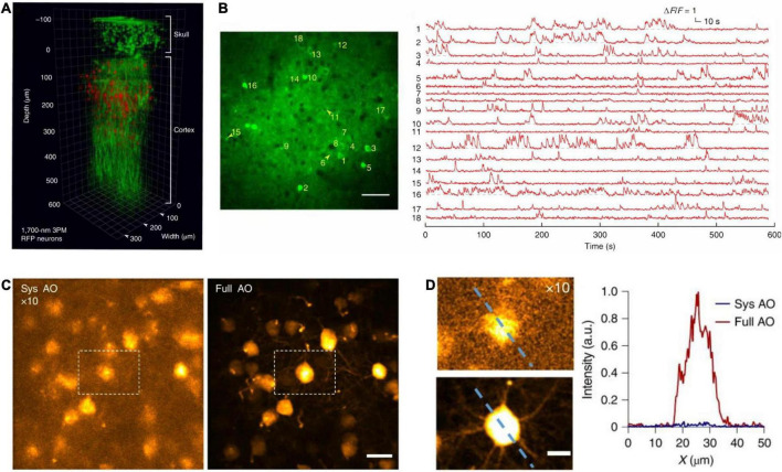
FIGURE 8.
In vivo cortical 3P imaging through the intact skull. (A) 3D reconstruction of CaMKII-positive neurons (∼140 μm below the cortical surface) labeled with GCaMP6s in a transgenic mouse. Images were captured by 1,320 nm 3PM through an intact skull of ∼100 μm thickness (red, fluorescence; green, THG). (B) Imaging site for through-skull activity recording in an awake, GCaMP6s-labeled transgenic mouse. The recording site was ∼275 μm beneath the dura, and the FOV was 320 μm× 320 μm (256 × 256 pixels per frame). Scale bar, 50 μm. (A,B) Were adapted with permission from Wang T. et al. (2018). (C) Maximum intensity projection of stack images (at the depth of 657–747 μm) of pyramidal neurons without (left) and with full AO correction (right). Scale bar, 20 μm. (D) Zoom in neurons in white dashed box in panel (C) (left). Scale bar, 10 μm. Signal intensity along the dashed line on left panel (right). a.u., arbitrary units. (C,D) Were adapted with permission from Ref. Qin et al. (2022).

