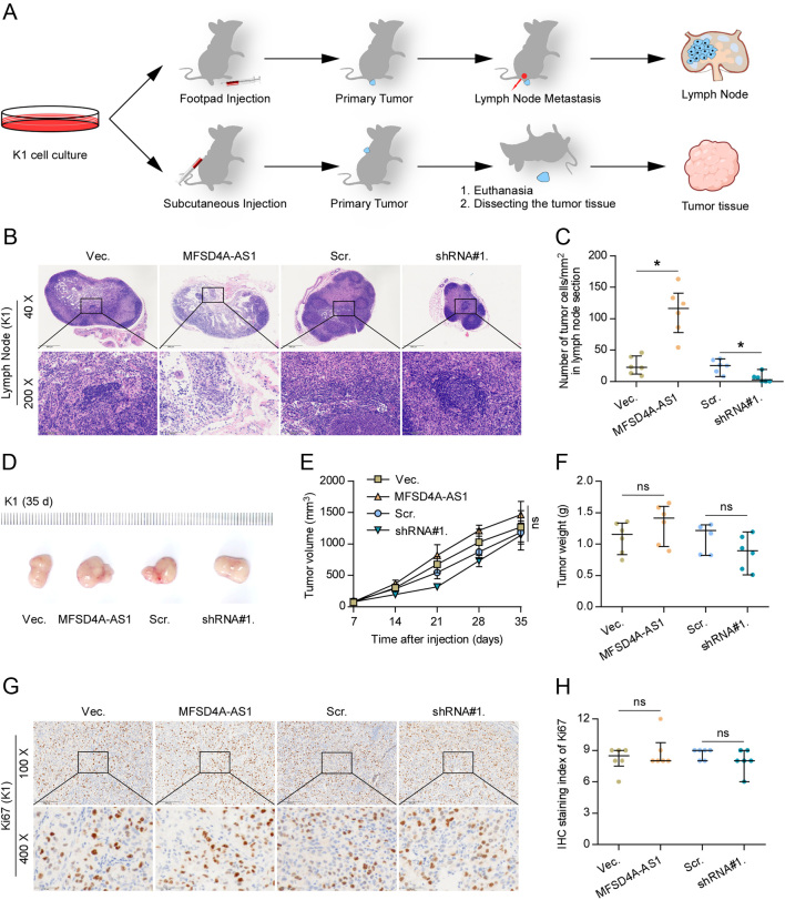
Figure 2.
MFSD4A-AS1 promotes lymphatic metastasis of PTC cells in vivo. (A) Schematic model of lymphatic metastasis (upper panel) and s.c. injection (upper panel) model in vivo. (B) H & E staining analysis of tumors in lymph nodes from the indicated mice group. (C) The number of tumor cells in the tumor areas of lymph nodes from the indicated mice group. *P < 0.05. (D) The representative tumors formed by the indicated K1 cells in each mice group (n = 6, each group). (E) The effect of MFSD4A-AS1 on the tumor volumes in the indicated mice groups from the seventh day at 7 days interval after inoculation of 5 × 10⁶ cells. Data presented are the mean ± s.d. n.s. indicates no significant difference. (F) The effect of MFSD4A-AS1 on the tumor weights in the indicated mice groups after inoculation of 5 × 10⁶ cells. n.s. indicates no significant difference. (G) The representative images of Ki-67 staining in the tumor tissues from the indicated mice groups. (H) The Ki-67 staining scores in the tumor tissues from the indicated mice groups. n.s. indicates no significant difference.

 This work is licensed under a