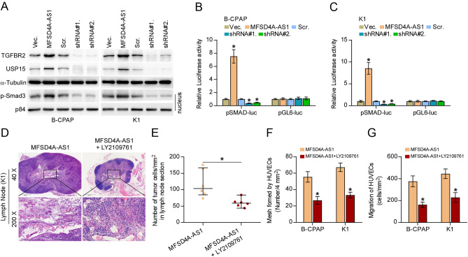
Figure 8.
MFSD4A-AS1 promotes lymphatic metastasis, invasion and migration of PTC cells partially by activating TGF-β signaling. (A) Western blotting analysis of the effect of overexpression or silencing MFSD4A-AS1 on TGFBR2, USP15 and nuclear translocation of phosphorylated Smad3 (p-Smad3) in the indicated PTC cells. α-Tubulin and p84 served as the cytoplasmic and nuclear loading control, respectively. (B and C) The effect of overexpression or silencing MFSD4A-AS1 on TGF-β/Smad-responsive luciferase reporter in the indicated cells. Error bars represent the mean ± s.d. of three independent experiments. *P < 0.05. (D) H & E staining analysis of tumors in lymph nodes from the indicated mice group. (E) The number of tumor cells in the tumor areas of lymph nodes from the indicated mice group. *P < 0.05. (F) The effect of TGF-β inhibitor LY2109761 in MFSD4A-AS1 overexpression PTC cells on tube formation ability of HUVECs in tube formation assay. Each bar represents the mean values ± s.d. of three independent experiments. *P < 0.05. (G) The effect of TGF-β inhibitor LY2109761 in MFSD4A-AS1 overexpression PTC cells on migration ability of HUVECs in the Transwell assay. Each bar represents the mean values ± s.d. of three independent experiments. *P < 0.05.

 This work is licensed under a