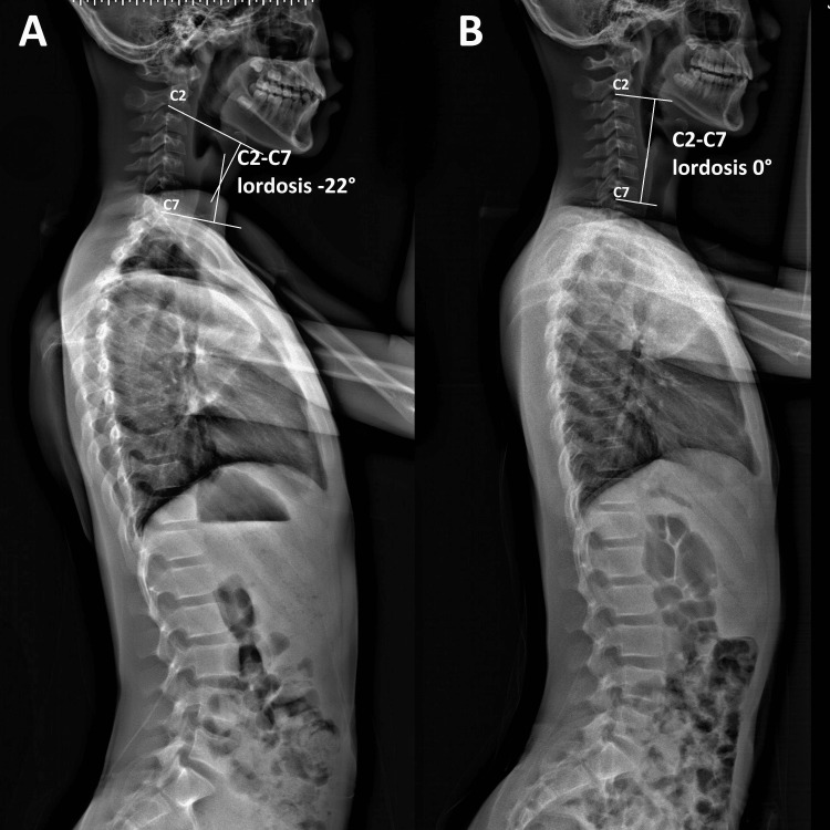
Figure 3. Full-spine radiographs (sagittal view) .
(A) Pre-treatment radiographs revealed reversed cervical lordosis. Using the Cobb angle (white lines), the C2-C7 spine’s global curvature was initially measure as –22°, indicating a reversed cervical lordosis. (B) At a 3-month follow-up, the cervical lordosis was corrected by 22° (0° vs. –22°) using the C2-C7 spine Cobb angle.

