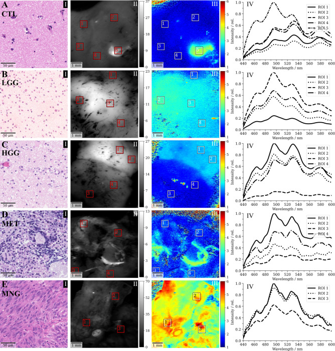
Figure 3.
Selected clinical cases of (A) non-tumorous control (CTL) tissue, (B) a WHO grade II diffuse astrocytoma (low-grade, LGG), (C) a glioblasatoma (high-grade, HGG), (D) a bronchial carcinoma metastasized to the cerebellum and (E) a meningothelial meningioma (MNG). (I) shows a representative haematoxylin and eosin (H&E) stain of the respective sample, (II) the demodulated fluorescence intensity in mVRMS, (III) the flavin fluorescence lifetime in ns (500-580 nm) and (IV) the spectroscopic measurements corresponding to the regions indicated in III.

