
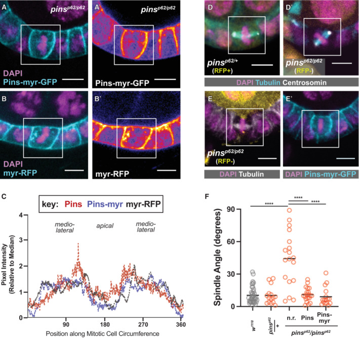
Figure 3. Pins does not require a Pins‐specific cue to relocalize at mitosis.
-
A–CPins‐myr‐GFP and myr‐RFP show a similar localization pattern to Pins:Tom during mitosis. Representative pictures are shown (A, B); a heatmap lookup table (A', B′) emphasizes the relative enrichments of Pins‐myr‐GFP and myr‐RFP along the cortex. Quantification (Pins‐myr: 4 cells, myr‐RFP: 2 cells) (C).
-
D–FRandom spindle orientation in pins p62 /pins p62 null mutant follicle cells is rescued by the expression of full‐length Pins or Pins‐myr‐GFP. Representative pictures (D, E) and quantification (F) are shown. Scale bars = 5 microns. Statistical significance in spindle orientation was determined using the Mann–Whitney test. ****P < 0.0001.