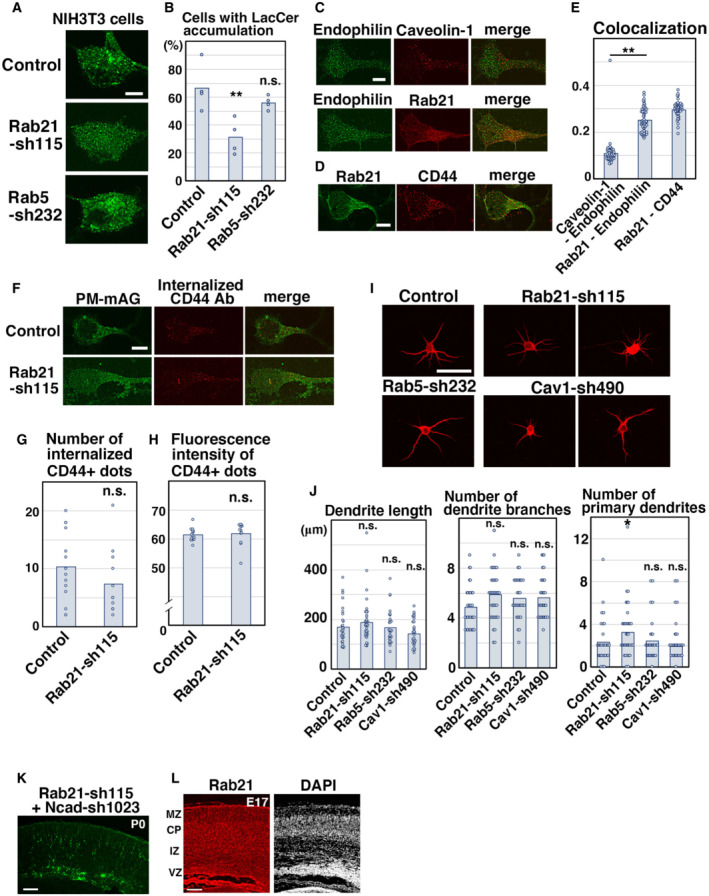
Figure EV4. Rab21 is required for the uptake of LacCer, but not CD44 internalization.

-
A, BNIH3T3 fibroblasts were transfected with the indicated plasmids and treated with BODIPY‐LacCer (LacCer) for 30 min before fixation. The images are obtained with high‐resolution microscopy (Nikon). The graph in (B) shows the ratio of cells with perinuclear accumulation of LacCer. Each score represents the mean with the individual points. Control: n = 4 biological replicates, Rab21‐sh115: n = 4 biological replicates, Rab5‐sh232: n = 4 biological replicates.
-
C–EPrimary cortical neurons from E15 cerebral cortices were incubated for 2 days in vitro and immunostained with the indicated antibodies. The images are obtained with high‐resolution microscopy (Nikon). The graph in (E) shows the Pearson's correlation coefficient between Endophilin and Rab21 or caveolin‐1 or between Rab21 and CD44. Each score represents the mean with the individual points. Caveolin‐1—Endophilin: n = 43 cells, Rab21—Endophilin: n = 53 cells, Rab21—CD44: n = 45 cells.
-
F–HPrimary cortical neurons from E15 cerebral cortices were transfected with the indicated plasmids plus pCAG‐PM‐mAG1, incubated for 2 days in vitro and subjected to CD44 antibody feeding assay. The graphs in (G) and (H) show the number of the internalized CD44‐positive dots and its total fluorescence intensity per cell. Each score represents the mean with the individual points. Control: n = 15 cells (G and H), Rab21‐sh115: n = 12 cells (G and H).
-
I, JPrimary cortical neurons from E15 cerebral cortices were transfected with the indicated plasmids plus pCAG‐EGFP, incubated for 8 days in vitro and stained with MAP2ab, a marker for dendrites. The graphs in (J) show the dendrite length, dendrite branch number and the number of primary dendrites. Each score represents the mean with the individual points. Control: n = 31 cells, Rab21‐sh115: n = 38 cells, Rab5‐sh232: n = 31 cells, Cav1‐sh490: n = 31 cells.
-
KCerebral cortex at P0, electroporated with the indicated plasmids plus pCAG‐EGFP at E14.
-
LFrozen sections of E17 cerebral cortex immunostained with anti‐Rab21 antibody and DAPI.
Data information: (B) Significance was determined by one‐way ANOVA with post hoc Dunnett and Tukey–Kramer. **Less than the critical value at 1%, *less than the critical value at 5%, n.s.: no significant differences. (E) Significance was determined by one‐way ANOVA with post hoc Tukey–Kramer. **Less than the critical value at 1%. (G, H) Significance was determined by Welch's t‐test (G: P = 0.1942) or Mann–Whitney's U test (H: P = 0.2225). n.s.: no significant differences. (J) Significance compared to control was determined by one‐way ANOVA with post hoc Dunnett. Significant difference was observed between control and Rab21‐sh115 in the number of primary dendrites. *Less than the critical value at 5%, n.s.: no significant differences. Scale bars: 5 μm in (A, C, D, F), 10 μm in (I), 150 μm in (K), 100 μm in (L).
