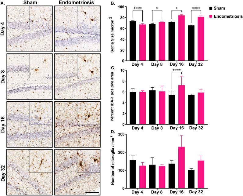
Fig. 5.
Microglial measurements in the hippocampus of sham controls and endometriotic mice. A Representative immunohistochemistry images of IBA1 in sham controls and endometriotic mice at various timepoints, the black bar in the lower right image is equal to 150 and 50 µm for the images and the insets, respectively. B Endometriotic mice had larger soma size on average than sham controls on days 8, 16, and 32. However, the soma size of the endometriotic mice was smaller on day 4 than sham controls. C Endometriotic mice also had increased IBA1 expression on day 16. D No difference in the microglia numbers was observed between the sham control and endometriotic mice. Values represent mean ± standard error mean (SEM), n = 5–6 mice/timepoint. The asterisks indicate significant differences between groups, *(p < 0.05) and ****(p < 0.0001)

