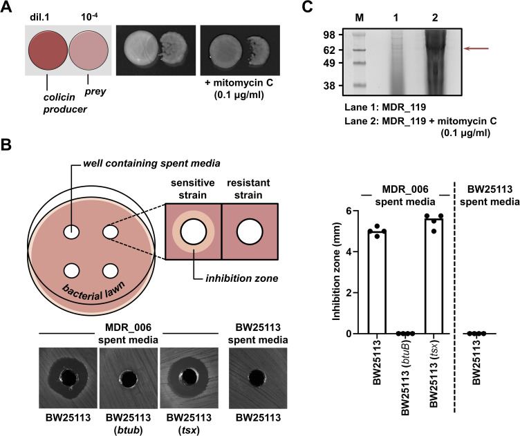
Figure 3.
Colicinogenic plasmids identified in CRE isolates are active in vitro. (A) MDR_006 kills other E. coli bacteria. MDR_006 was spotted onto LB agar (at 0.8% w/v) without or with mitomycin C (at a final concentration of 0.1 μg mL−1) next to the E. coli K-12 strain BW25113,22 with each strain at a different dilution (1 vs 10−4, respectively). Plates were incubated at 37 °C overnight. The left image shows a schematic representation of the competition conditions. (B) pCol006 harbored by MDR006 encodes a Colicin E1-like toxin. LB agar (at 1.5% w/v) plates were swabbed with E. coli BW25113, its btuB or tsx mutant (as obtained from the Keio collection),22 and four wells were carved into each plate. Wells were filled with culture supernatant (spent media) either produced by MDR_006 or by E. coli BW25113 after overnight growth in the presence of 0.1 μg mL−1 mitomycin C. This panel contains a schematic representation of the growth inhibition assay (left; top), representative images of the obtained growth inhibition zones for all strain-spent media combinations (left; bottom), and quantification of the growth inhibition zones (right). (C) MDR_119 releases cloacin toxin when it is exposed to DNA damage. The panel shows an SDS-PAGE analysis of TCA-precipitated culture supernatants of MDR_119 after overnight growth in the absence (lane 1) or presence (lane 2) of mitomycin C (at a final concentration of 0.5 μg mL−1). Molecular weight markers (M) are on the left, the red arrow indicates the position of the bands originating from the cloacin toxin.

