Figure 2.
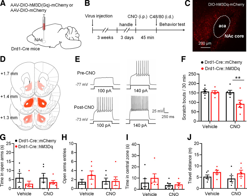
Chemogenetic activation of Drd1-MSNs in the NAc core decreases C48/80-induced scratching behaviors. A, Schematic illustration of virus injection site in the NAc. The AAV-DIO-hM3D(Gq)-mCherry was used for conditional activation of Drd1-MSNs in Drd1-Cre mice. B, Schematic diagram of the time line for the experiment. C, Representative brain section shows the expression of AAV-DIO-hM3Dq-mCherry in the NAc core. aca, anterior commissure, anterior part. D, Viral spread of AAV-DIO-hM3D(Gq)-mCherry and AAV-DIO-mCherry in Drd1-cre mice. Each mouse is represented by one translucent outline, and outlines are overlapped. E, Sample traces shows the injected currents (+100 and +140 pA)-evoked membrane voltage responses recorded in hM3Dq-expressing Drd1-MSNs before and after CNO (5 μm) application. Notably, CNO incubating leads a relatively sustained depolarization on the RMP and increases the firing frequency in hM3Dq-expressing cells. F, Chemogenetic activation of Drd1-MSNs in the NAc core decreases the scratching behaviors in mice treated with C48/80 (F(interaction 1,10) = 8.282, p = 0.0164; F(drug 1,10) = 10.16, p = 0.0097; F(virus 1,10) = 5.298, **p = 0.041). G, The EPMT shows that chemogentic activation of Drd1-MSNs in the NAc core does not change the time spent in open arms in mice treated with C48/80 (F(interaction 1,10) = 0.03761, p = 0.8501; F(drug 1,10) = 0.04718, p = 0.8324; F(virus 1,10) = 2.805, p = 0.1249). H, Same as G for summary of the open arms entry (F(interaction 1,10) = 1.462, p = 0.2544; F(drug 1,10) = 0.8845, p = 0.3691; F(virus 1,10) = 0.6358, p = 0.4438). I, The OFT shows that chemogenetic activation of Drd1-MSNs in the NAc core does not change the time spent in central zone in mice treated with C48/80 (F(interaction 1,10) = 0.3991, p = 0.5417; F(drug 1,10) = 2.062, p = 0.1816; F(virus 1,10) = 0.7666, p = 0.4018). J, Same as I for summary of the travel distance (F(interaction 1,10) = 0.01651, p = 0.9003; F(drug 1,10) = 0.5096, p = 0.4916; F(virus 1,10) = 3.048, p = 0.1114).

