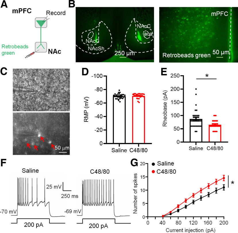
Figure 6.
C48/80 increases the excitability of mPFC-to-NAc projection pyramidal neurons. A, Schematic diagram for retrobeads green injection and patch-clamp recording in brain slices. B, Representative coronal brain slices show retrobeads injection sites in the NAc core and retrobeads green-positive neurons (green) in the mPFC. aca, anterior commissure, anterior part; NAcC, NAc core; NAcSh, NAc shell. C, Images show the electrophysiological recording on retrobeads-labeled pyramidal neurons in the mPFC. Red arrows marked the retrobeads-positive neurons. D, Summary bar graph and scatter plot for RMP of green retrobeads-positive PFC-projecting to NAc pyramidal neurons (n = 27–29 neurons, p = 0.3098, U = 329, Mann–Whitney U test). E, Same as D for rheobase of pyramidal neurons (n = 27–29 neurons, *p = 0.0110, U = 244, Mann–Whitney U test). F, Sample traces of membrane voltage responses recorded from green retrobeads-positive PFC-projecting to NAc pyramidal neurons from saline-treated or C48/80-treated mice. G, Summarized data show the number of evoked spikes in the groups as indicated in F (n = 27–29 neurons, F(interaction 8,400) = 4.816, p < 0.0001; F(treatments 1,50) = 6.576, *p = 0.0134, two-way ANOVA with repeated measure).

