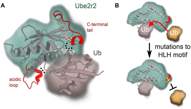
Figure 5. Ube2r2 positions the donor ubiquitin and regulates its activity.

(A) The acidic loop and C-terminal domain (both in red ribbons, interactions indicated by dotted lines) of Ube2r2 can help position ubiquitin in the closed conformation (PDB 6NYO). (B) The helix-loop-helix motif (HLH, in red) on Ube2r2 appears to be important for positioning of the acceptor ubiquitin. Mutations (yellow) at this site result in a decrease in acceptor ubiquitin binding and activity.
