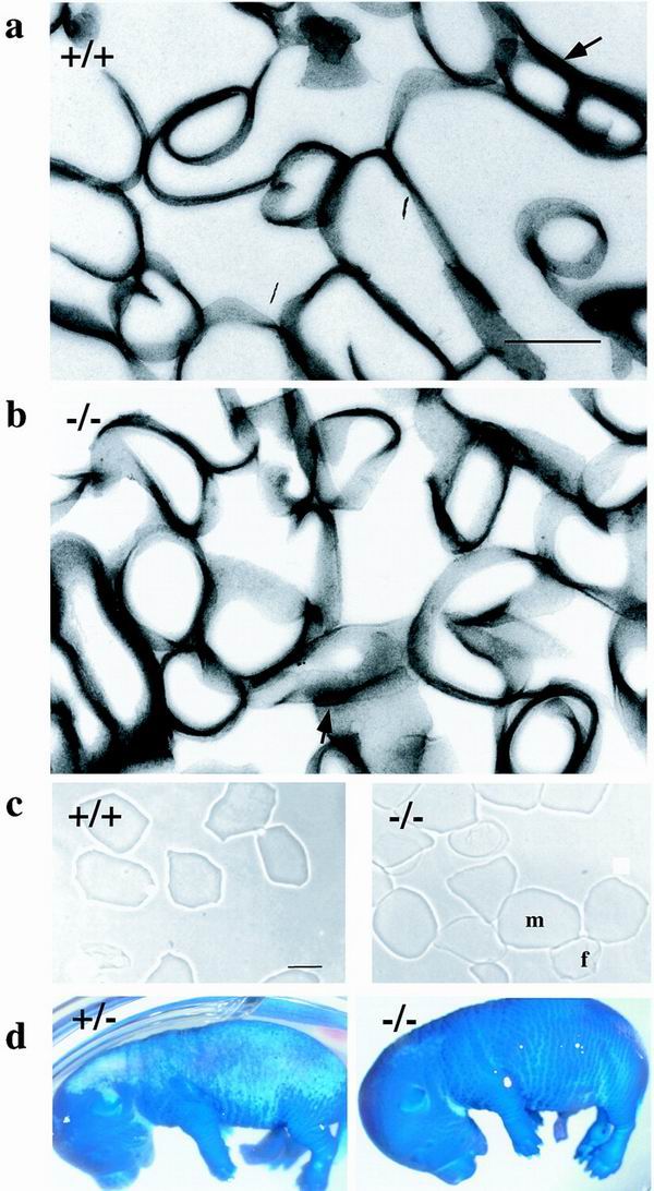
FIG. 3.
Barrier acquisition and cornified envelope morphology. (a and b) Transmission electron microscopy of isolated CE from +/+ (a) and −/− (b) epidermis. Arrows indicate residual desmosomes. (c) Cornified envelopes isolated from 2-day-old wild-type or envoplakin knockout mice. f, fragile envelope; m, mature envelope. (d) E16.5 heterozygous (+/−) and envoplakin null homozygous (−/−) embryos stained with toluidine blue. Dark blue areas of the skin have not yet formed the epidermal barrier. Bar, 500 nm (a and b) or 25 μm (c).

