Figure 1.

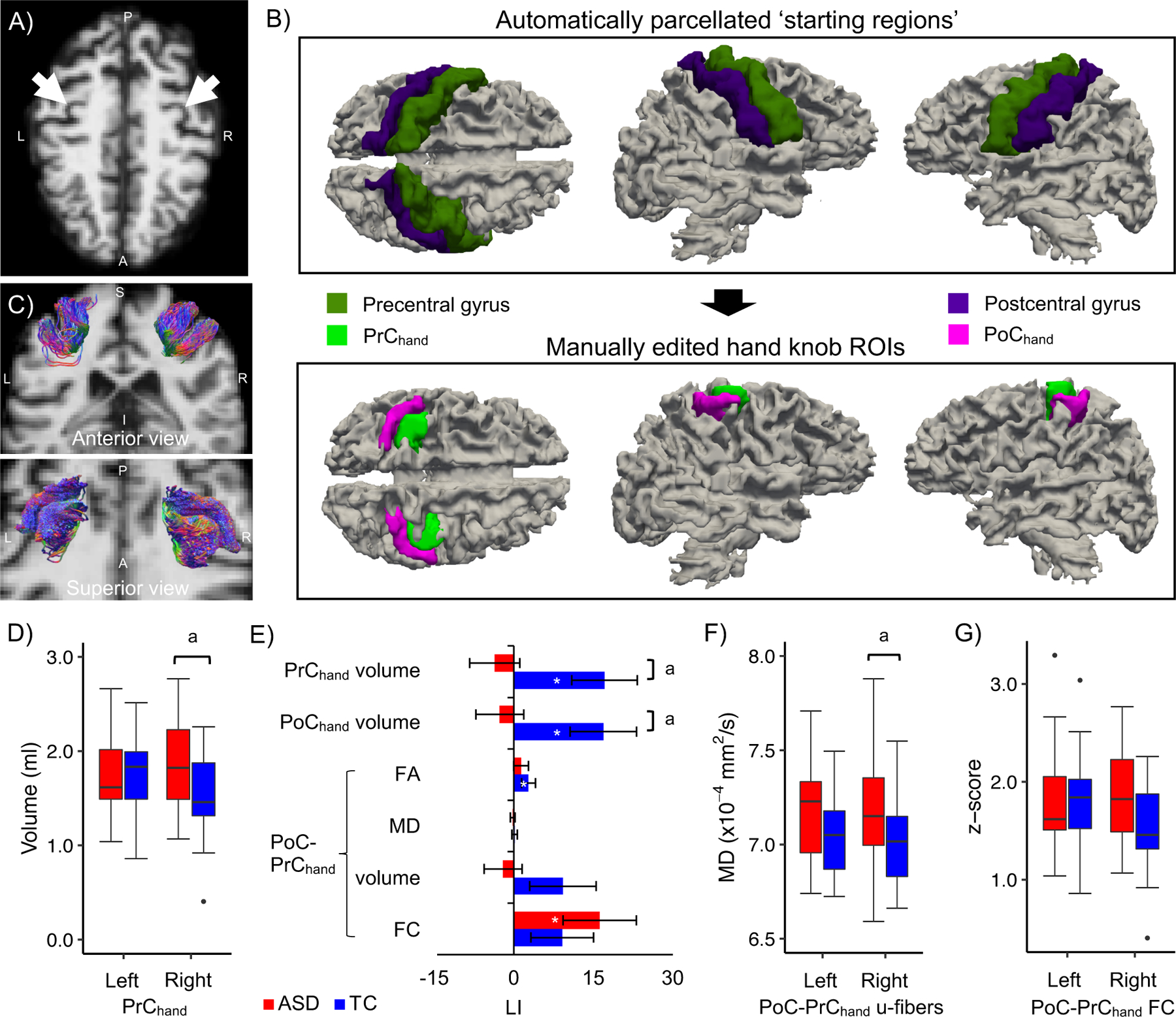
A-C) Identification and delineation of the hand knob regions and u-fibers. A) The typical omega-shaped landmark (white arrows) used to identify the hand knob in the axial plane of a representative participant with ASD (aged 49 years). B) The automatically parcellated pre and postcentral regions (upper row) were manually edited to define the pre (PrChand) and postcentral (PoChand) hand knob regions of interest (lower row). C) Streamline tractography of the u-fiber tracts connecting ipsilateral PoChand and PrChand from the same participant. D-G) Alterations in hand knob morphometry, laterality and PoC-PrC connectivity in middle-aged adults with ASD. The plots show D) increased right PrChand volume, E) reduced laterality of PrChand and PoChand knob volume (LI of other hand knob measures are also shown), and F) increased mean diffusivity (MD) in the right PoC-PrChand u-fiber tract in the ASD group. G) Medium size group effects were also observed for left PoC-PrChand u-fiber MD (increased in ASD) and right PoC-PrChand FC (decreased in ASD). a q<.10 significance level; * LI measures that differed from zero (p<.05, uncorrected). Standard errors of the means are shown in the bar plot.
