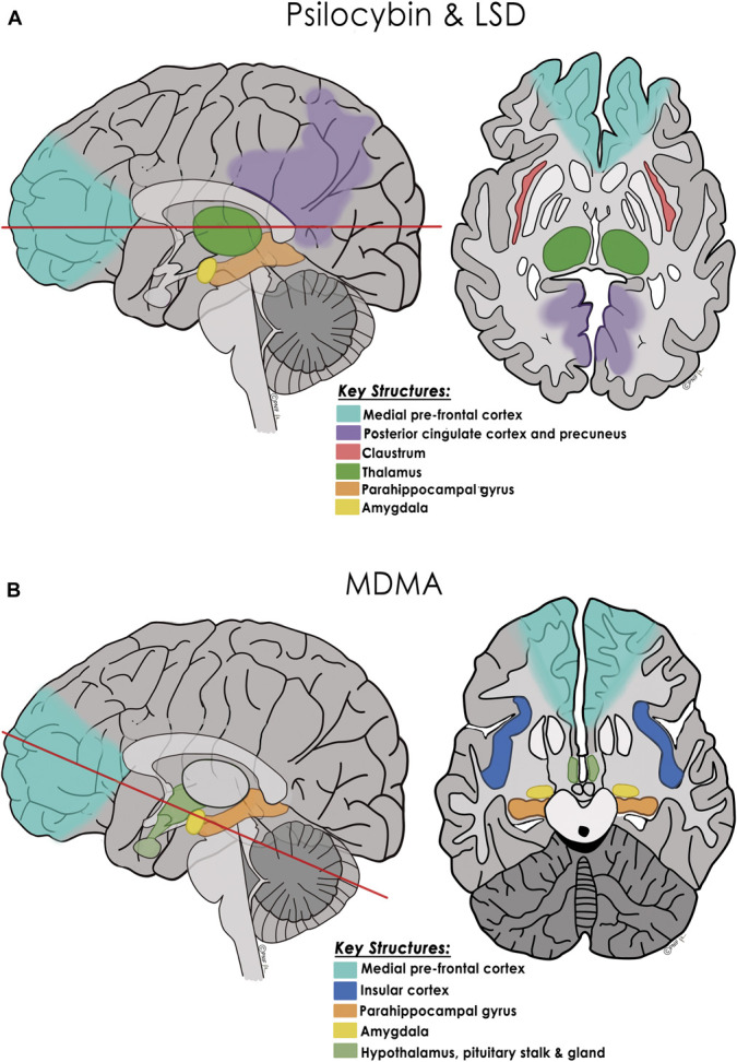
FIGURE 4.

Proposed key neural structures and mechanism of action of the classic psychedelics and MDMA. A, Psilocybin and LSD: Based on functional neuroimaging studies, sagittal and axial drawings show major brain regions altered by psilocybin and LSD (predominantly 5-HT2A receptor agonists): pyramidal neurons in the medial PFC, the DMN (including the posterior cingulate cortex and precuneus), and other networks such as the salience network, the thalamus, claustrum, parahippocampal gyri, and amygdala (red line in sagittal image denotes axial cut level).12,16,59-61,64 Three functional models have been proposed for how the serotonergic psychedelics alter these cortical and subcortical structures and their regional connectivity. While they differ as to the predominance of “top-down” or “bottom-up” loosening, all 3 models propose that the normally highly filtered flow of sensory information and access to autobiographical information is dampened or partially disrupted in the acute psychedelic state. This more connected state is proposed to lead to the commonly experienced auditory-visual synesthesias, visual hallucinations, alterations of space and time, alterations of one's sense of self, and in many patients, a mystical experience.59,60,63 The CSTC model proposes that activation of medial prefrontal cortex 5-HT2AR–containing neurons projecting to the striatum disrupts thalamocortical gating leading to reduced thalamic filtering of sensory information and increased signaling to the cerebral cortex, triggering synesthesias, cognitive, and emotional changes, and in some patients, ego dissolution.16,61,63,64 The “relaxed beliefs under psychedelics” model proposes that psilocybin and LSD increase information flow from the hippocampus and parahippocampal gyrus to higher cortical areas, including the DMN, which relax high-level prior beliefs of one's sense of self, ego, and social identity.60 This temporary disruption of the normal “top-down” constraint imposed over neural networks and sensory processing, along with favorable changes in amygdala responsiveness, is believed to halt or dampen ruminative thoughts and potentiate a more functionally connected and flexible brain.5,65 The cortico-claustro-cortical model, focuses on the claustrum, situated between the insula and the putamen with a high density of 5-HT2A receptors and considered critical in maintaining cortical networks including the DMN and frontoparietal network.59 Based on fMRI data, psilocybin acutely decreases claustrum connectivity to auditory and other cortical areas with resultant alterations in perception and attention; this decreased cognitive control correlates with measures of a “mystical experience.”12,59 B, MDMA: Based on neuroimaging studies, sagittal and axial drawings show major brain regions and neurohormonal circuits altered by MDMA including connections between the medial PFC, the amygdala, parahippocampal gyri, and insula, as well as the hypothalamic-pituitary axis.34 (Red line in sagittal image denotes axial cut level.) While MDMA does not seem to cause as much increased connectivity between neural networks as do psilocybin and LSD, MDMA-induced release of oxytocin (facilitated by serotonin efflux) affects connectivity between the medial PFC and amygdala, dampening amygdalar activation which may contribute to prosocial effects and memory reconsolidation. MDMA-induced cortisol release may also affect the amygdala and hippocampus, allowing extinction learning by facilitating emotional engagement despite revisiting fear and anxiety related to a traumatic event.34 CSTC, cortico-striatial-thalamo-cortical; DMN, default mode network; LSD, lysergic acid diethylamide; MDMA, 3,4-methylenedioxymethamphetamine. © Pacific Neuroscience Institute Foundation, 2022. Used with permission.
