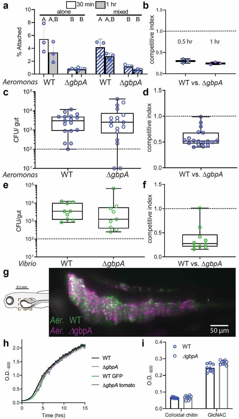
Figure 3.

Colonization of the zebrafish intestine by A. veronii and V. cholerae does not require gbpA. (a) Binding of WT and ∆gbpa A. veronii to chitin beads, quantified as the percent of total bacteria, after either 0.5 hr (white bars) or 1 hour (gray bars) of incubation. Bacterial strains were added to chitin beads alone (solid bars) or mixed with the other strain (striped bars). Boxplot whiskers represent range. Groups with different letter designations are statistically different with a p value of < 0.05 whereas groups with the same letter are not significantly different. (b) Competitive index of ∆gbpa versus WT A. veronii recovered from chitin beads. (c) A. veronii CFUs recovered at 6 dpf following inoculation of GF zebrafish with individual strains at 4 dpf. (d) Competitive index of ∆gbpa versus WT A. veronii recovered at 6 dpf following co-inoculation of GF zebrafish with the two strains at 4 dpf. (e) V. cholerae CFUs recovered at 6 dpf following inoculation of GF zebrafish with individual strains at 4 dpf. (f) Competitive index of ∆gbpa versus WT V. cholerae strains recovered at 6 dpf fish following co-inoculation of GF zebrafish with the two strains at 4 dpf. (g) Light sheet micrograph of zebrafish intestine colonized with WT (green) and ∆gbpa (purple) A. veronii in the proximal intestinal region indicated in the schematic. (h) Growth curves measuring OD600 for each A. veronii strain grown in TSB. (i) Final OD600 measurement for WT and ∆gbpa strains grown on colloidal chitin and GlcNAc.
