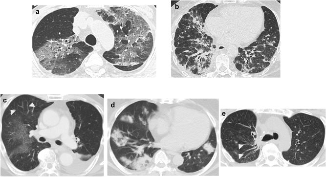
Figure 2.
Drug-related pneumonitis showing new diffuse ground-glass opacities, consolidation, and traction bronchiectasis, indicative of a diffuse alveolar damage pattern (a). Drug-related pneumonitis showing new ground-glass opacities, irregular reticular opacities, and irregular reticular opacities with predominant lower lung involvement, indicative of the nonspecific interstitial pneumonia pattern (b). Drug-related pneumonitis showing new wide areas of faint ground-glass opacities with some patchy nodular lesions (arrowheads), indicative of the hypersensitivity pneumonitis pattern (c). Drug-related pneumonitis showing new ground-glass opacities and consolidations with multifocal distribution, indicative of the organizing pneumonia pattern (d). Drug-related pneumonitis showing new focal opacity areas (arrowheads). Lesions disappear only with withdrawal of drug therapy (not shown here), with features compatible with the simple pulmonary eosinophilia pattern (e).

