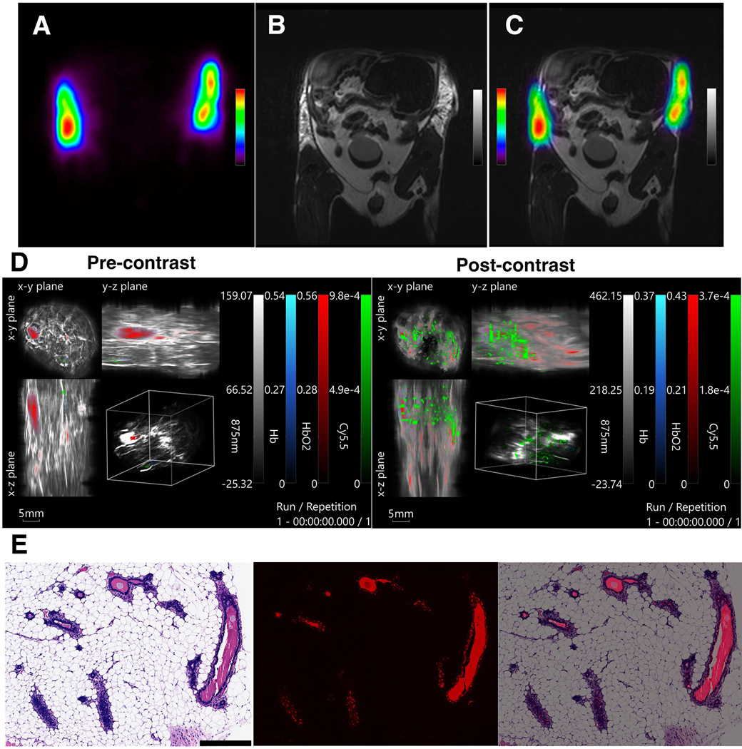
Figure 5.

In vivo imaging of intraductal injections of the fully conjugated contrast agent, radiolabeled with Cu-64, imaged with A) PET and B) MRI, and the C) overlay of the two modalities. D) Photoacoustic imaging (left – pre-contrast; right – post-contrast). E) Histology of the excised ductal tree tissue. Left: H&E stain of the ductal epithelial cells after injection with the contrast agent. Middle: Fluorescence of the agent accumulated in the ductal epithelial cells, imaged in Cy5.5 channel; Right: the merged images.
