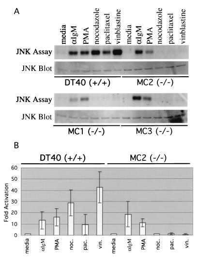
FIG. 2.
JNK activation in DT40 cells after stimulation with microtubule-disrupting agents is abolished in mekk1−/− cell lines MC1, MC2, and MC3. (A) DT40, MC1, MC2, and MC3 cells were incubated in DT40 medium alone (30 min), anti-IgM (15 min), 50 ng of PMA per ml–250 ng of ionomycin per ml (30 min), 1 μg of nocodazole (noc.) per ml (30 min), 1 μM paclitaxel (pac.) (30 min), and 1 μM vinblastine sulfate (vin.) (30 min) at 37°C. Cells were subsequently lysed in modified radioimmunoprecipitation assay buffer, and 300 μg of total protein from each extract was used in a JNK in vitro kinase assay as described in Materials and Methods. For the JNK Western blot assay, 25 μg of total protein from each extract was subjected to SDS-PAGE. The filter was incubated with an anti-JNK (C17) antibody. (B) Three separate JNK in vitro kinase assays were performed, and results were quantitated as described in Materials and Methods. Results are expressed as fold activation over untreated cells. Average values are shown.

