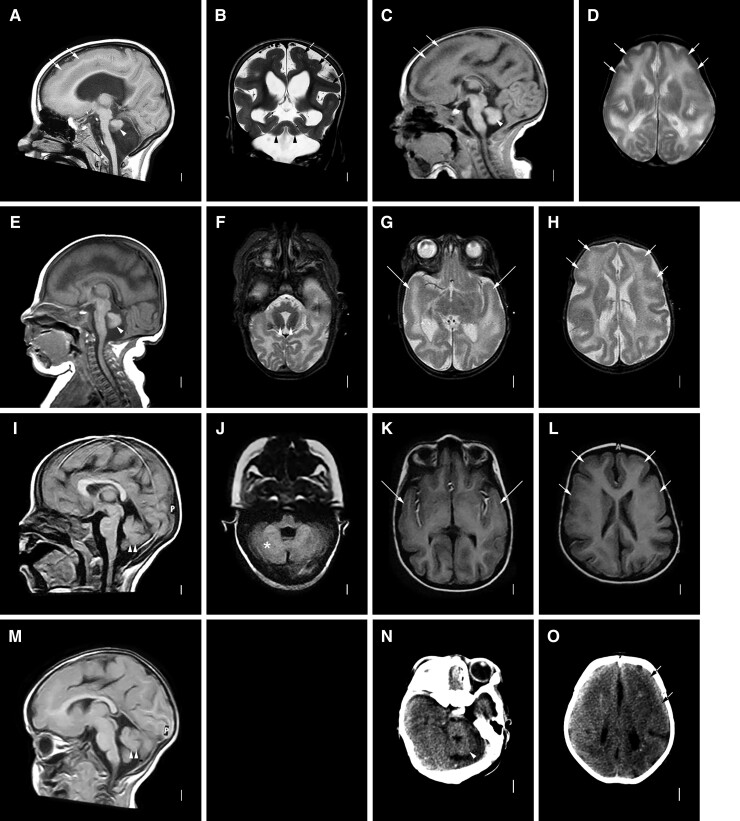
Figure 2.
Brain MRI showing LIS-CBLH with biallelic RELN variants. Brain imaging in six individuals with biallelic RELN-related LIS-CBLH. Midline sagittal MRIs in three individuals show an abnormally thin brainstem with flat pons and very small cerebellum with an afoliar surface (arrowheads in A, C and E). Midline sagittal images in two siblings with a less severe mutation show a normal brainstem and only moderately small cerebella with some foliation evident (I, M; double arrowheads point to the small cerebella). Axial (D, F–H, J–L) and coronal (B) images shows diffuse but frontal and temporal-predominant LIS with moderately thick 5–8 mm cortex. Arrows point to representative areas of pachygyria (mild LIS) only; the cortical malformation affects all brain regions. Low resolution head CT images in another child show the small cerebellum (arrowhead in N) and mild LIS (arrows in O). The imaging pattern seen in panels A–H appears similar to those shown in prior reports of individuals with biallelic mutations, while the pattern seen in panels I–M are less severe. Panels A and B are from subject LP95-137a1; C and D from LP95-137a2, E and H from LR14-063, I and L from LR17-413a1, M from LR17-413a3 and N and O from LP96-078.

