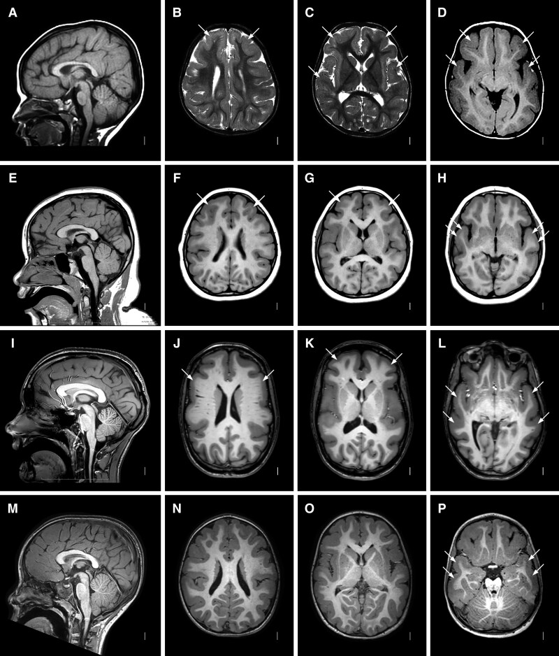
Figure 3.
Brain MRI showing frontotemporal LIS with monoallelic RELN variants. Images show the same pattern of LIS in four unrelated subjects including LR02-111a2 (A–D), LR21-446 (E–H), LR15-139 (I–L) and LR18-437 (M–P). Midline sagittal images (first column) show normal brainstem and cerebellum. Axial images at the level of the lateral ventricles (second column), basal ganglia (three column) and temporal lobes (fourth column) show diffuse mild LIS (pachygyria) with only moderately thick 5–8 mm cortex and consistent gradient with the malformation most severe in the frontal and temporal lobes and becoming less severe posteriorly. The posterior parietal and occipital regions appear mildly abnormal, although this is sometimes subtle. Arrows point to representative areas of pachygyria. The hippocampi also appear normal (not shown).

