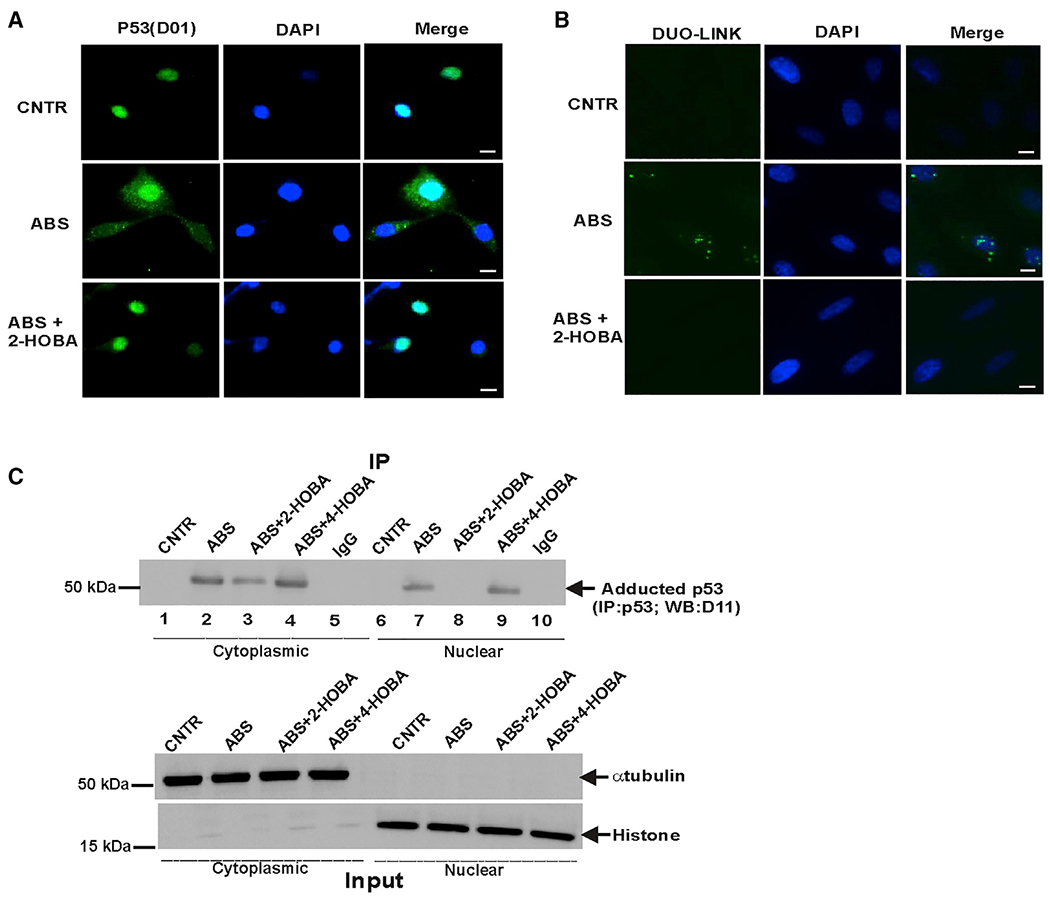
Figure 4. p53 adduction leads to p53 protein aggregation.

(A) Representative images of p53 protein aggregates in CP-A cells (scale bar, 5 μm). Immunofluorescence staining was performed with p53(D0-1) antibody in cells treated with ABS alone or in combination with 2-HOBA. 2-HOBA prevented aggregation of p53 protein (n = 3).
(B) Representative images of Duolink PLA in CP-A cells treated with ABS. PLA was performed using D0-1 and D11: E-tag primary antibodies (scale bar, 5 μm). The isoLG-p53 adducts were detected in large cellular aggregates in cells treated with ABS. The adduct-specific PLA signals were not observed in cells treated with 2-HOBA (n = 3).
(C) p53 protein adduction was analyzed in the cytoplasmic and nuclear fractions of CP-A cells after ABS treatment for 8 h. Equal amount of total protein (10 μg) was loaded in each well. Bottom panel shows experimental inputs.
