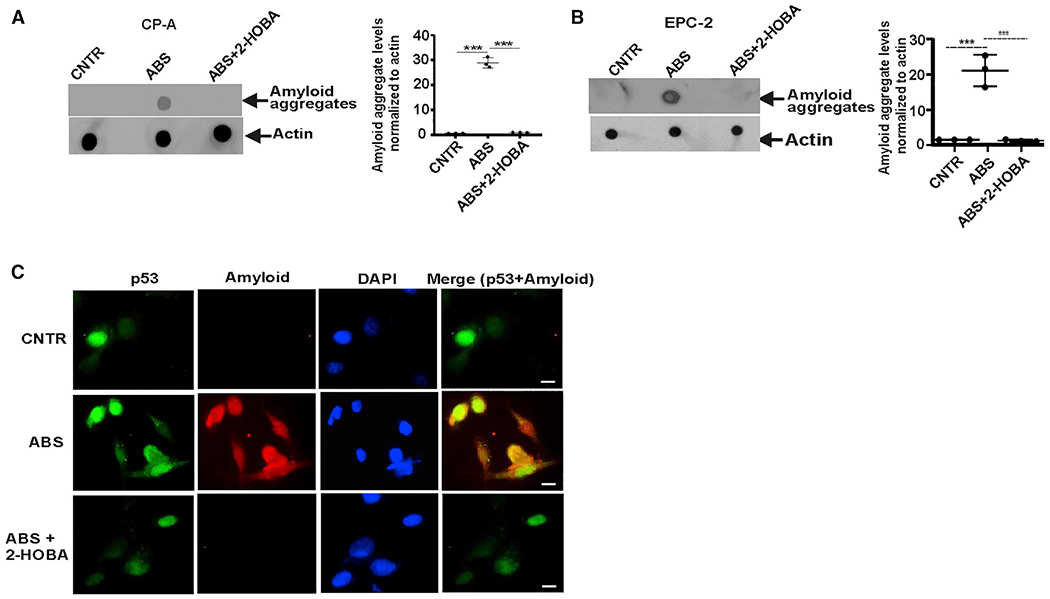
Figure 6. IsoLGs form amyloid-like p53 aggregates.

(A) Dot blot analysis of CP-A cell extracts using anti-amyloid OC antibody. Protein aggregation levels were found to be higher in CP-A cells treated with ABS than in control untreated cells (***p < 0.001; n = 3; Tukey’s multiple comparison). 2-HOBA prevented amyloid aggregation (***p < 0.001; n = 3; Tukey’s multiple comparison).
(B) The same as (A) but the formation of amyloid aggregates was analyzed in EPC-2 cells.
(C) Representative images of co-localization of p53 protein with amyloid fibrils detected with anti-amyloid OC antibody in CP-A cells (n = 3) (scale bar, 5 μm). All results are expressed as mean ± SD.
