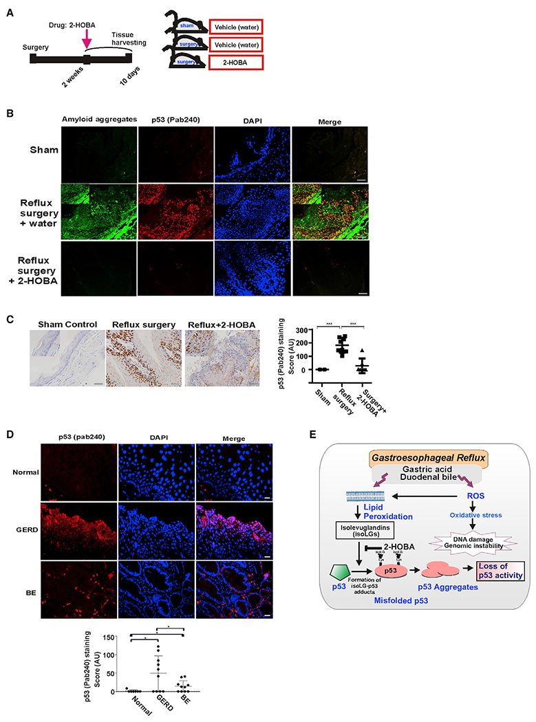
Figure 7. IsoLGs induce misfolding of p53 protein in vivo.

(A) The schematic representation of animal experiment.
(B) Representative images of p53 protein co-localization with amyloid fibrils detected using amyloid OC antibody in murine esophageal tissues. Mice received 2-HOBA dissolved in water (1 g/L) 2 weeks after surgery. Control mice received vehicle (water). Esophageal tissues were collected after drug treatments for 10 days and analyzed for misfolding of p53 protein. Co-localization of p53 and amyloid staining was observed in mice with reflux surgery. The staining was not found in mice treated with 2-HOBA. Inset: high magnification image. Scale bar, 10 μm.
(C) Representative immunohistochemical staining of murine esophageal tissues with p53 (PAb 240) antibody. Insets show high magnification images. Graph shows the IHC scores (sham control versus reflux mice, ***p < 0.001; reflux mice versus reflux mice treated with 2-HOBA, ***p < 0.001; Tukey’s multiple comparison). Mice with sham surgery were used as a control. Scale bar, 10 μm.
(D) Representative images of p53 protein misfolding in the human esophagus. The human esophageal tissues from healthy subjects, GERD and BE patients were analyzed using p53 (PAb 240) antibody. Misfolded p53 protein was observed in the esophagus of GERD and BE patients, but not in normal (no GERD) esophageal epithelium (scale bar, 5 μm). Graph shows the immunofluorescence scores (normal versus GERD, *p < 0.01; normal versus BE, *p < 0.01).
(E) Graphical representation of p53 protein regulation by isoLGs. All results are expressed as mean ± SD.
