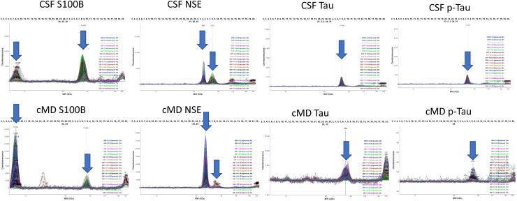
FIG. 1.
Distribution of immunoreactivities of S100B, NSE, Tau, and p-Tau in CSF and bECF samples. Blue arrows point to the immunoreactive peaks that were quantified. bECF, brain extracellular fluid; CSF, cerebrospinal fluid; NSE, neuron-specific enolase; p-Tau, phosphorylated Tau; S100B, S100 calcium-binding protein B.

