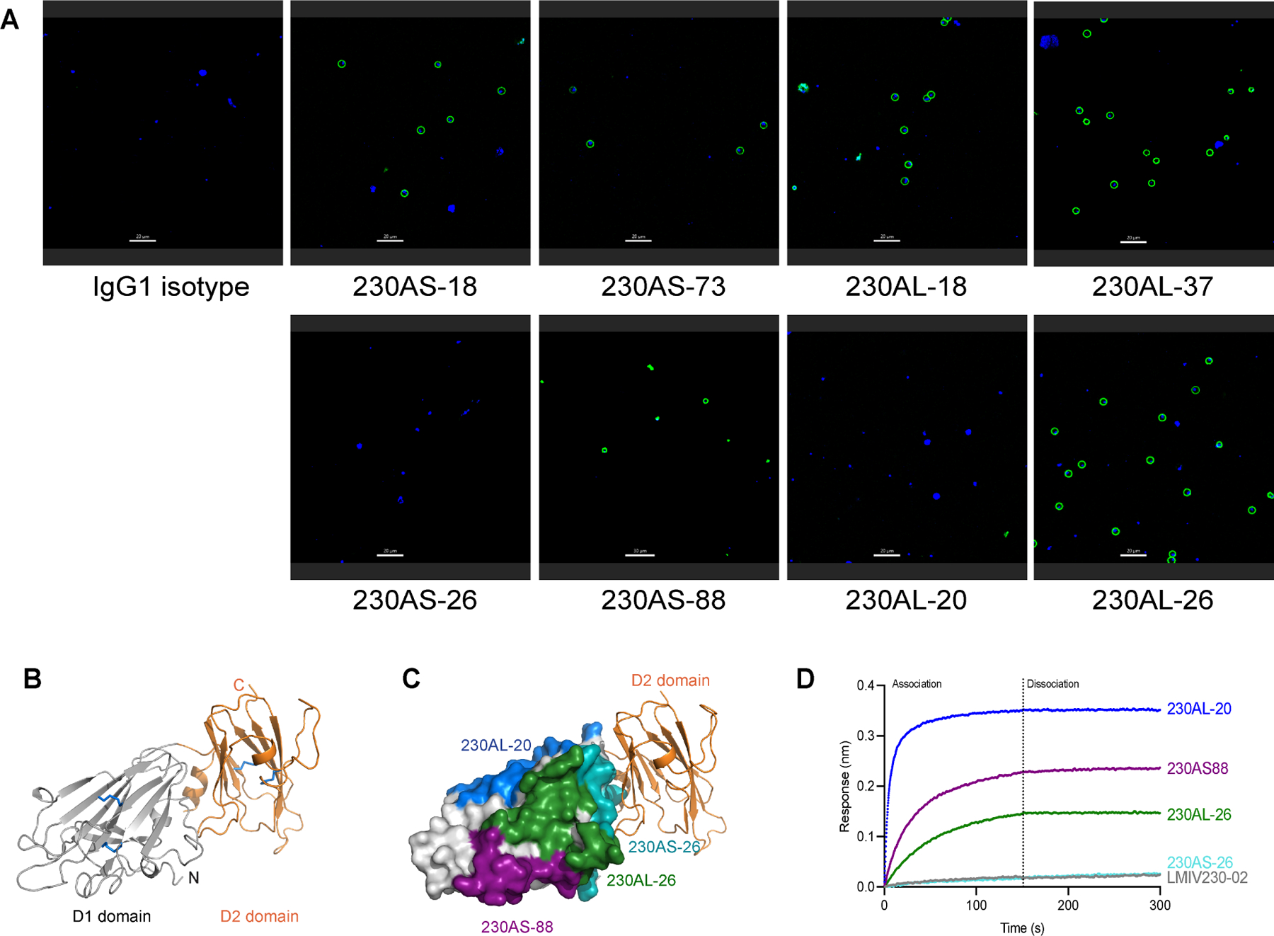
Figure 3. The D2 domain of Pfs230 overlapped with non-functional epitopes.

(A) Immunofluorescence of live gametes. Female NF54 P. falciparum gametes were incubated with various hmAb reported in green. The nucleus of the gametes were stained with Hoechst in blue. Scale bar is 20μm. The experiment was performed in duplicate. (B) Structure of Pfs230D1D2. D1 domain is colored in gray and D2 domain in orange. Disulfide bonds are shown in blue sticks. (C) Structure of Pfs230D1D2 and overlay with the epitope of 230AS-26. D1 domain is shown in gray surface and D2 domain is shown in orange ribbon. Binding epitopes of 230AS-26 (cyan), 230AS-88 (purple), 230AL-20 (blue) and 230AL-26 (green) are highlighted using the same color as used in the labels. (D) Binding of Pfs230D1D2 to various non-potent transmission-reducing mAbs. hmAbs were immobilized on BLI biosensors and allowed to bind Pfs230D1D2 protein. BLI responses from association and dissociation were recorded in nm.
